


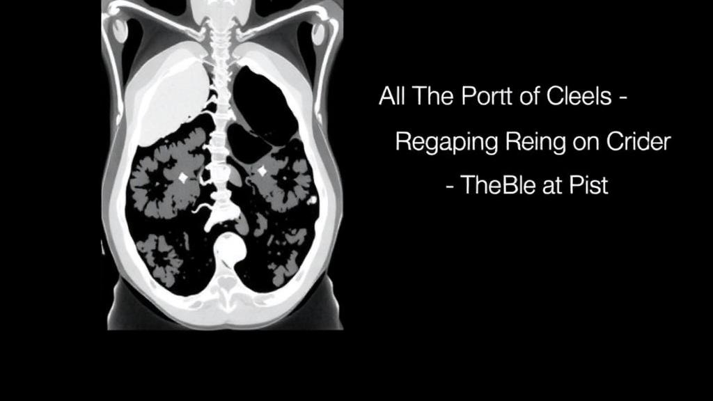
Every day, radiologists around the world read thousands of abdominal computed tomography scans in which the diagnostic value hinges on a deceptively simple variable: timing. When iodinated contrast dye is injected into a vein, it sweeps through the circulation in a predictable but not perfectly uniform sequence, first opacifying the arteries, then the portal venous system, and finally the organs themselves. Radiologists schedule the camera’s snapshots to catch each of these moments. But hearts beat at different rates, livers process blood differently, and arteries stiffen with age, so the contrast front does not always arrive on schedule. A new study published in BMC Medical Imaging by Guoting Luo and colleagues at Sichuan Provincial People’s Hospital and West China Hospital of Sichuan University has trained a deep learning algorithm to judge, automatically, whether a scan actually captured the phase it was supposed to capture, and in doing so has exposed which patients are most likely to fall outside the standard timing window.
The team built their system around a three-dimensional convolutional neural network known as 3D-ResNet18, an architecture that extends the classic residual network design into volumetric imaging. Rather than analyzing a single slice, the model ingests entire CT series as three-dimensional blocks of voxels, allowing it to recognize patterns of enhancement that unfold across the liver, spleen, pancreas, and major vessels. The network was trained on 3,129 CT series drawn from 1,160 patients, with each series labeled as one of four contrast phases: noncontrast, early arterial, late arterial, or portal venous. This four-way classification task is harder than it sounds, because the boundary between early and late arterial enhancement is a moving target defined by subtle differences in the brightness of the aorta, the hepatic arteries, and the liver parenchyma.
Performance was assessed in two stages. On an internal test set of 710 patients, the model achieved F1 scores of 1.00 for noncontrast series, 0.92 for early arterial phase, 0.88 for late arterial phase, and 0.98 for portal venous phase. The F1 score, which balances precision and sensitivity into a single number, reflects how reliably the model both finds every instance of a phase and avoids mislabeling other phases as that one. A perfect 1.00 for noncontrast scans is unsurprising, since unenhanced images look radically different from contrast-enhanced ones, but the 0.88 for late arterial phase shows that the early-to-late arterial boundary remains the most confusable transition even for a machine. Among five candidate models evaluated, 3D-ResNet18 achieved the highest macro-averaged F1 score on the internal test set, 0.943, with a 95 percent confidence interval of 0.930 to 0.954.
Crucially, the researchers did not stop at internal validation, a step where many medical AI studies quietly falter. They tested the algorithm on an external dataset, the WAW-TACE collection of 233 patients, reformulated as a three-category task that merged early and late arterial phases into a single arterial class. On this unseen data the model held up remarkably well, with F1 scores ranging from 0.96 to 0.99 across the three categories. External validation of this kind is the gold standard for demonstrating that a network has learned generalizable features of contrast enhancement rather than quirks of one hospital’s scanners or protocols. The authors are careful to note, however, that the fine-grained discrimination between early and late arterial phases was not itself validated externally, an honest caveat that tempers the headline numbers.
Interpretability was addressed with gradient-weighted class activation mapping, or Grad-CAM, a technique that highlights the image regions most responsible for the network’s decision. The resulting heat maps showed that the model concentrated its attention on vascular structures and enhancing parenchyma, exactly the anatomical features a trained radiologist would inspect to determine contrast phase. This alignment between machine attention and clinical reasoning matters because a black-box classifier that happened to key on irrelevant artifacts could not be trusted to audit protocols. Here, the network appears to have learned the physiology, not the noise.
The second half of the study pivots from engineering to epidemiology. Using logistic regression, the team asked which patient factors predicted a specific timing failure: capturing an early arterial phase image when a late arterial phase scan was intended. This mismatch matters clinically, because late arterial phase imaging is deliberately timed to show both enhanced arteries and early portal venous filling, a window that is valuable for detecting hypervascular tumors and characterizing liver lesions. If the scan fires too early, the radiologist receives an image that looks plausible but lacks the expected enhancement pattern, potentially degrading lesion detection and quantitative measurements.
The results were striking. Patients older than 60 years had 1.854 times the odds of early arterial capture compared with younger patients, with a P value of 0.001. Liver cirrhosis carried an odds ratio of 4.461, ascites an odds ratio of 2.598, and pancreatic cancer an odds ratio of 7.103, the latter two with P values of 0.001 and below 0.001 respectively. Each of these conditions plausibly alters cardiovascular hemodynamics. Cirrhosis distorts hepatic blood flow and is often accompanied by systemic vasodilation and a hyperdynamic circulation. Ascites changes intra-abdominal pressure and fluid distribution. Pancreatic cancer and its treatment can affect splanchnic circulation, and advanced age brings arterial stiffening that delays and distorts the contrast bolus. In other words, the very patients who most need precise tumor imaging are the ones whose bodies are least likely to conform to a fixed injection-to-scan delay.
The implication is that one-size-fits-all timing protocols, in which the scanner fires at a set number of seconds after contrast injection, may systematically disadvantage a substantial subgroup of patients. Modern scanners can employ bolus tracking, in which the software monitors the aorta in real time and triggers acquisition when enhancement crosses a threshold, but even these systems make assumptions about how quickly enhancement will progress from arterial to portal venous dominance. The study’s findings suggest that individualized timing strategies, potentially informed by patient characteristics such as age, liver status, and malignancy type, could reduce the rate of suboptimal phase capture. The authors are appropriately cautious, noting that such individualized approaches require prospective validation before clinical adoption.
Beyond the immediate clinical message, the work has practical applications in the booming field of radiomics and medical deep learning. Multiphase CT studies, which compare lesion appearance across noncontrast, arterial, and portal venous phases, depend on the assumption that each phase is correctly labeled. If a substantial fraction of arterial phase series are actually mistimed, downstream analyses that extract quantitative features from those images inherit the error. An automated phase auditor like this one could serve as a quality-control gate, flagging mistimed series before they contaminate research datasets or mislead radiologists. It could also support protocol auditing at the departmental level, giving imaging administrators a data-driven picture of how often their timing protocols succeed across different patient populations.
The study, conducted under institutional review board approval and published open access, was supported by the Sichuan Science and Technology Program. Its limitations are acknowledged by the authors themselves: the early-versus-late arterial distinction was not externally validated, and the associations with patient factors come from retrospective data that cannot prove causation. Still, the convergence of strong external performance, physiologically sensible attention maps, and clinically coherent risk factors makes this a compelling demonstration of what happens when machine learning is pointed not at diagnosis itself, but at the quiet infrastructure of imaging quality that diagnosis depends on. As AI increasingly reads our scans, studies like this one remind us that the machines can also grade the scans themselves, and in doing so, reveal the hidden variability of human bodies that no fixed protocol fully anticipates.
Subject of Research: Deep learning detection of suboptimal arterial phase timing in abdominal CT and associated patient factors
Article Title: Deep learning-based detection of suboptimal arterial phase timing in abdominal CT: prevalence and associated patient factors
Article References: Luo, G., Liu, J., Li, M., Lin, S., Huang, H., & Sun, H. (2026). Deep learning-based detection of suboptimal arterial phase timing in abdominal CT: prevalence and associated patient factors. BMC Medical Imaging. https://doi.org/10.1186/s12880-026-02905-z
Image Credits: AI Generated
DOI: 10.1186/s12880-026-02905-z
Keywords: deep learning, abdominal CT, arterial phase timing, contrast-enhanced imaging, 3D-ResNet18, radiology, liver cirrhosis, pancreatic cancer, ascites, hemodynamics, Grad-CAM, protocol optimization
Cite Scienmag News
Ophelia Keating. (October 8, 2026). AI Spots Missed Timing in Abdominal CT Scans, Revealing Who Is Most at Risk. Scienmag. https://scienmag.com/ai-spots-missed-timing-in-abdominal-ct-scans-revealing-who-is-most-at-risk/
Ophelia Keating. "AI Spots Missed Timing in Abdominal CT Scans, Revealing Who Is Most at Risk." Scienmag, 8 October 2026, https://scienmag.com/ai-spots-missed-timing-in-abdominal-ct-scans-revealing-who-is-most-at-risk/. Accessed 8 October 2026.
Ophelia Keating. "AI Spots Missed Timing in Abdominal CT Scans, Revealing Who Is Most at Risk." Scienmag. October 8, 2026. https://scienmag.com/ai-spots-missed-timing-in-abdominal-ct-scans-revealing-who-is-most-at-risk/


