


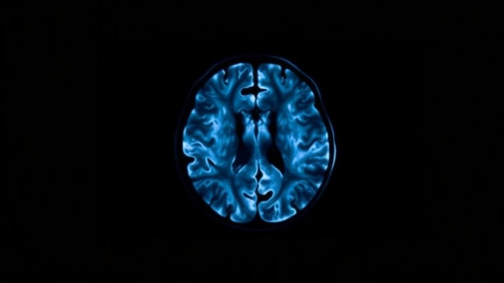
A single brain scan can quietly reveal whether a person with fuzzy memory is standing at the edge of Alzheimer’s disease, and a new real-world study now shows that the answer holds up no matter how the scan is read. In research published in the European Journal of Nuclear Medicine and Molecular Imaging, a team from Clínica Universidad de Navarra in Pamplona, Spain, followed 145 patients with amnestic mild cognitive impairment for up to eight years and found that a positive amyloid PET scan strongly predicted who would go on to develop Alzheimer’s dementia, regardless of whether the images were interpreted by expert eyes, with computerized statistical support, or by hard numbers alone.
Amnestic mild cognitive impairment, or aMCI, is widely regarded as the prodromal phase of Alzheimer’s disease, a stage where memory complaints are measurable but daily function is largely preserved. Not everyone in this stage progresses, however, and clinicians have long sought tools to separate those carrying underlying beta-amyloid pathology from those whose memory problems stem from other causes. Amyloid positron emission tomography answers that question by using fluorine-18-labelled radiotracers that bind to beta-amyloid deposits in the cortex, making the molecular signature of the disease visible in a living brain. Patients whose scans show pathological cortical retention are known to be at substantially higher risk of clinical decline, which is why amyloid PET has become central to diagnosis and, increasingly, to the selection of patients for anti-amyloid therapies.
Yet the way a scan is interpreted has always been a point of debate. Visual reading, the current clinical standard, depends on the trained eye of a nuclear medicine physician comparing grey and white matter contrast against tracer-specific criteria. It is fast and well validated, but it is inherently subjective, and borderline scans with low or intermediate tracer retention can divide even experienced readers. To reduce that subjectivity, imaging platforms now offer semi-quantitative support: standardized uptake value ratios, or SUVRs, that condense tracer signal in composite cortical regions into a single number compared against validated thresholds, and z-score maps that highlight voxels deviating from a normative database of age-matched healthy controls. The updated Appropriate Use Criteria from the Alzheimer’s Association and the Society of Nuclear Medicine and Molecular Imaging endorse these tools as complements to visual interpretation in selected scenarios, but whether they genuinely improve reproducibility or prognostic power beyond expert reading has remained uncertain.
The Navarra team set out to answer that question directly in a routine memory clinic setting. Between October 2013 and March 2021, they enrolled 145 patients diagnosed with aMCI after comprehensive neurological, neuropsychological, laboratory and MRI workups, excluding anyone over 85, those with depression, abnormal laboratory findings, or alternative brain injuries such as lacunar infarcts. Three different fluorinated amyloid tracers were used in clinical practice: florbetapir in 79 patients, flutemetamol in 46, and florbetaben in 20, all acquired on the same Biograph mCT PET/CT scanner with time-of-flight and point-spread-function reconstruction. Patients were then followed clinically until January 2022, with progression defined by the National Institute on Ageing–Alzheimer’s Association 2011 criteria for Alzheimer’s disease dementia.
Two certified nuclear medicine physicians, one with more than 30 years of experience and one with five, independently read every scan twice: first with conventional structured visual interpretation, and then with assistance from the syngo.via MI Neuro Cortical Analysis software, which overlays voxel-wise z-score statistics and SUVR values onto the images. As a third method, scans were classified purely by SUVR thresholds validated in prior literature, above 1.17 for florbetapir, above 1.35 for florbetaben and above 0.60 for flutemetamol. Agreement between and within readers was quantified with Cohen’s kappa, a statistic in which values above 0.81 conventionally signal excellent agreement.
The reliability results were striking. Inter-observer agreement reached a kappa of 0.877 for structured visual reading and 0.904 for assisted interpretation, while intra-observer consistency between the two approaches was 0.860 for the senior reader and 0.892 for the junior one. When consensus ratings were established with the assisted method, agreement climbed to 0.936, near the statistical ceiling. Reader 1 classified 94 scans as amyloid-positive on visual reading and 101 with assistance; Reader 2 classified 96 and 97 respectively. In other words, experienced readers agreed with themselves and with each other at a level that leaves very little room for chance, confirming that standardized visual interpretation of amyloid PET is far more reproducible than its subjective reputation might suggest.
Reproducibility, however, is only half the story; the real question is whether a positive scan predicts what happens next. Using Cox proportional hazards models adjusted for age, sex and education, the researchers found that amyloid positivity significantly increased the risk of conversion to Alzheimer’s dementia under all three interpretation methods. Assisted visual interpretation produced the strongest signal: PET-positive patients had a 3.23-fold higher risk of progression than PET-negative patients, with a hazard ratio of 3.23 and a 95 percent confidence interval of 1.94 to 5.38. SUVR quantification yielded a hazard ratio of 2.19, and conventional visual reading, 1.83. Kaplan–Meier survival curves separated significantly between positive and negative groups for every method, with log-rank tests all significant below the 0.001 level. Over the follow-up period, 75 patients converted to Alzheimer’s dementia, while 41 remained clinically stable.
The most intriguing findings came from the scans where readers disagreed. Eighteen of the 145 studies, 12.4 percent, showed discordance across at least one interpretative condition, and in this small, exploratory subgroup the hierarchy of methods flipped. When clinical progression was used as the pragmatic reference standard, conventional visual reading outperformed assisted interpretation for both readers: the senior reader correctly classified 9 of 18 discordant cases visually versus only 2 with assistance, and SUVR binary classification matched clinical outcome in 11 of 18 cases, the best of all. A detailed re-evaluation showed the disagreements were not random. Most involved florbetapir scans, and cortical atrophy was flagged as a contributing factor in four, while other cases reflected reduced image quality, subtle or borderline tracer uptake, or atypical regional distribution. Disagreement, in short, clustered in technically and biologically challenging images rather than revealing a flaw in either human or machine reading.
The authors are careful to frame these subgroup findings as descriptive rather than definitive, given the small sample, and they acknowledge that the higher hazard ratio for assisted interpretation at the cohort level may seem at odds with its weaker performance on the hardest cases. Their resolution is nuanced: quantitative tools appear to sharpen prognostic stratification across a whole clinic population, likely by nudging borderline calls toward the correct side, but they offer little incremental value for expert readers confronting genuinely ambiguous scans. This mirrors prior multicentre work showing that disagreement in amyloid PET concentrates in scans with low or borderline amyloid burden, and earlier trials demonstrating that trained visual readers detect beta-amyloid pathology with high sensitivity and specificity, including autopsy-validated correlations between florbetapir signal and plaque density.
The implications reach well beyond the reading room. As anti-amyloid immunotherapies move into routine practice, amyloid PET is no longer an academic exercise; it gates treatment decisions and, increasingly, therapeutic monitoring, with a growing trend toward reporting Centiloid values that harmonize quantification across tracers. The product characteristics of all three radiotracers already recommend quantitative analysis as a complement to visual assessment, and this study supports that framework while adding a caution: software overlays and machine-learning classifiers now emerging for automated amyloid classification should be treated as decision-support tools, not replacements for expert oversight, especially when atrophy, motion, or borderline uptake complicate the picture. The study’s limitations, including the absence of autopsy confirmation, the use of tracer-specific thresholds rather than Centiloid conversion, and a single institutional reconstruction protocol, leave room for refinement. But the core message is clear and reassuring: in the real world, a positive amyloid PET scan in a patient with amnestic mild cognitive impairment is a robust, method-independent warning of Alzheimer’s disease on the horizon, and the trained human eye remains an indispensable part of reading it.
Subject of Research: Prognostic value of amyloid PET interpretation methods for predicting Alzheimer's disease conversion in amnestic mild cognitive impairment
Article Title: Prognostic value of Amyloid PET in Real-World Amnestic MCI: Influence of reader experience and interpretation method
Article References: Echeveste, B., Guillén, E. F., Prieto, E., Hirschmüller, K. C. E., Riverol, M., & Arbizu, J. (2026). Prognostic value of Amyloid PET in Real-World Amnestic MCI: Influence of reader experience and interpretation method. European Journal of Nuclear Medicine and Molecular Imaging. https://doi.org/10.1007/s00259-026-08159-9
Image Credits: AI Generated
DOI: 10.1007/s00259-026-08159-9
Keywords: Alzheimer's disease, amnestic mild cognitive impairment, amyloid PET, visual interpretation, SUVR, z-score maps, inter-observer agreement, reader expertise, prognosis, nuclear medicine, biomarkers, dementia
Cite Scienmag News
Cassandra Pierce. (October 3, 2026). Amyloid PET Scans Predict Alzheimer’s Risk No Matter How Experts Read Them. Scienmag. https://scienmag.com/amyloid-pet-scans-predict-alzheimers-risk-no-matter-how-experts-read-them/
Cassandra Pierce. "Amyloid PET Scans Predict Alzheimer’s Risk No Matter How Experts Read Them." Scienmag, 3 October 2026, https://scienmag.com/amyloid-pet-scans-predict-alzheimers-risk-no-matter-how-experts-read-them/. Accessed 3 October 2026.
Cassandra Pierce. "Amyloid PET Scans Predict Alzheimer’s Risk No Matter How Experts Read Them." Scienmag. October 3, 2026. https://scienmag.com/amyloid-pet-scans-predict-alzheimers-risk-no-matter-how-experts-read-them/


