


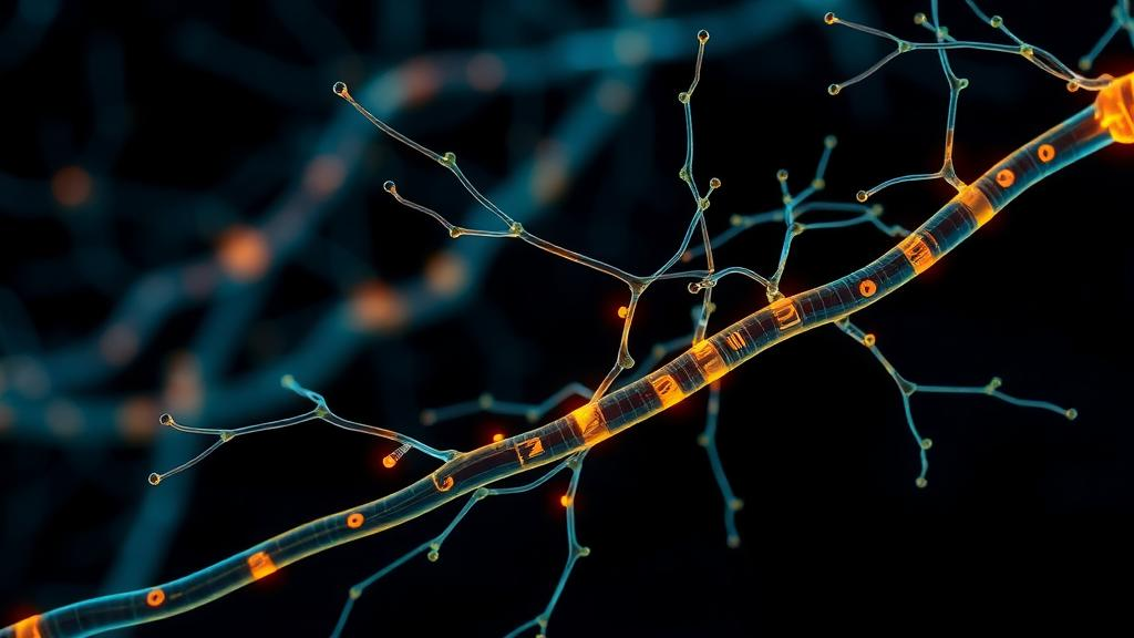
Every thought, movement, and sensation depends on electrical signals racing through the nervous system at precisely calibrated speeds, and one of the most important determinants of that speed is a fatty wrapping called myelin. In a study published this month in PLOS Biology, researchers at State University of New York Upstate Medical University report the identification of a key mechanism that helps determine the length of individual myelin segments, offering a new explanation for how the brain and spinal cord organize the intricate wiring of the nervous system. The work, led by senior author Marie Bechler, PhD, assistant professor of cell and developmental biology and of neuroscience and physiology, with recent PhD graduate Amanda R. Young as first author, points to a mechanosensitive protein called Piezo1 as a critical sensor that allows myelin-forming cells to measure the nerve fibers they envelop and adjust their output accordingly.
Myelin is produced in the central nervous system by specialized glial cells known as oligodendrocytes. These cells extend processes that wrap around nerve fibers, or axons, much like insulation around an electrical wire, forming compact sheaths that allow signals to travel efficiently through the brain and spinal cord. The analogy is more than decorative: just as a cable with damaged insulation loses current, an axon whose myelin is compromised loses the ability to conduct rapid, reliable impulses. Myelin sheaths are important to just about everything we do, and when they are damaged and lost, as occurs in multiple sclerosis, signaling between neurons becomes disrupted and the support needed to maintain neuronal health is lost, producing devastating symptoms related to body control, fatigue, vision, thinking, and movement.
One of the enduring puzzles in neurobiology has been that myelin is not uniform. The length of individual myelin segments varies throughout the nervous system, and those variations are not random. For decades, researchers have known that thicker nerve fibers tend to have longer myelin segments, a scaling relationship that helps tune conduction properties across the diverse repertoire of axons found in a mature nervous system. What has remained unclear is how the cells that produce myelin detect the size of a nerve fiber and use that information to determine how much myelin to make. The new study provides a direct answer to the first half of that question, identifying a molecular ruler of sorts embedded in the myelin-forming cells themselves.
That ruler is Piezo1, a protein best known in other contexts as a mechanically activated ion channel. According to the Upstate-led team, oligodendrocytes use Piezo1 to sense the diameter of the nerve fibers they are wrapping, and that mechanical information helps determine the length of each myelin segment they build. In other words, the geometry of the axon is translated, through a force-sensitive channel, into a decision about how far a given sheath should extend. This finding reframes myelination not simply as a biochemical program but as a process in which physical properties of the target axon are actively read and interpreted by the wrapping cell.
The temporal dimension of the discovery is equally significant. The researchers found that Piezo1 appears to play an especially important role during the early stages of myelin formation, when oligodendrocytes are actively building and extending the myelin sheath. This suggests that the window in which axon diameter is measured and translated into sheath length is the same window in which the fundamental architecture of a myelinated fiber is being established. Once a sheath has been laid down, its dimensions influence how quickly nerve signals travel along that fiber, so errors made during the construction phase could have lasting consequences for circuit performance.
Understanding how myelin sheath length is regulated matters well beyond developmental biology, because myelin is essential for efficient nerve signaling and healthy brain and spinal cord function. Damage to myelin is associated with conditions such as multiple sclerosis and other neurological disorders, and the mechanisms that govern sheath formation in the first place are likely to intersect with the mechanisms that determine whether damaged sheaths can be repaired. By identifying how the nervous system establishes precise patterns of myelin along its nerve fibers, the study provides a foundation for future investigations into myelin repair and regeneration, a goal that has proven difficult to achieve clinically despite decades of effort.
Senior author Marie Bechler framed the motivation for the work in terms of disease across the lifespan. “Numerous neurological conditions across our lifespan disrupt oligodendrocyte cells and the myelin sheaths they form,” she said. “Our research aims to understand the impact of these changes compared to the healthy nervous system as well as to find ways to promote myelin sheath growth in diseases where myelin is lost or damaged.” The statement underscores a central theme of modern glial biology: to repair myelin therapeutically, researchers first need a detailed blueprint of how healthy myelin is built, segment by segment, along every axon it insulates.
The technical achievement behind the study relied on specialized infrastructure. The research was conducted by a team of Upstate Medical University investigators in the Bechler lab, with part of the study assisted by the institution’s Electron Microscopy core, a facility whose high-resolution imaging capabilities are essential for quantifying structures as small and precisely organized as myelin sheaths and the axons beneath them. Measuring whether sheath lengths scale with axon diameter requires exactly this kind of careful anatomical reconstruction, and the involvement of a dedicated core facility reflects the meticulous, quantitative nature of the question being asked.
The publication itself, titled “Myelin sheath lengths in the central nervous system scale to axon diameter via oligodendroglial Piezo1,” appeared in PLOS Biology on 21 September 2026, carrying the DOI 10.1371/journal.pbio.3003992. The authors reported no conflicts of interest. Placement in a high-profile open-access journal signals that the finding is expected to interest a broad audience, from developmental neuroscientists studying how glial cells interpret their environment to clinicians searching for actionable targets in demyelinating disease. Because Piezo1 is a channel protein rather than an anonymous genetic factor, it offers a concrete molecular entry point for future experiments aimed at manipulating myelin formation.
For the field, the study closes a conceptual loop that has been open for decades. Axon diameter was known to predict sheath length; oligodendrocytes were known to be the cells that must somehow perform the prediction; and now a specific mechanosensitive molecule has been identified as the conduit through which axonal geometry informs myelination decisions. The broader implication is that the wiring of the nervous system is shaped not only by genetic instruction and neuronal activity but also by the physical dialogue between axons and the cells that insulate them. As research into myelin repair accelerates, mechanisms like the one uncovered at Upstate Medical University are likely to define the roadmap, showing where healthy construction can be mimicked and where damaged sheaths might one day be coaxed to regrow with the right dimensions, in the right places, restoring the fast and faithful signaling on which the entire nervous system depends.
Subject of Research: Mechanisms regulating myelin sheath length in the central nervous system
Article Title: Upstate researchers uncover clue to how the brain wires itself
Article References: Upstate researchers uncover clue to how the brain wires itself. (n.d.). Original publication
Image Credits: AI Generated
DOI: Not provided
Keywords: myelin, oligodendrocytes, Piezo1, axon diameter, PLOS Biology, multiple sclerosis, neuroscience, SUNY Upstate Medical University, myelination, demyelination, nerve signaling, glial cells
Cite Scienmag News
Cassandra Pierce. (September 30, 2026). Piezo1 protein helps the brain measure nerve fibers to build myelin of the right length. Scienmag. https://scienmag.com/piezo1-protein-helps-the-brain-measure-nerve-fibers-to-build-myelin-of-the-right-length/
Cassandra Pierce. "Piezo1 protein helps the brain measure nerve fibers to build myelin of the right length." Scienmag, 30 September 2026, https://scienmag.com/piezo1-protein-helps-the-brain-measure-nerve-fibers-to-build-myelin-of-the-right-length/. Accessed 30 September 2026.
Cassandra Pierce. "Piezo1 protein helps the brain measure nerve fibers to build myelin of the right length." Scienmag. September 30, 2026. https://scienmag.com/piezo1-protein-helps-the-brain-measure-nerve-fibers-to-build-myelin-of-the-right-length/


