


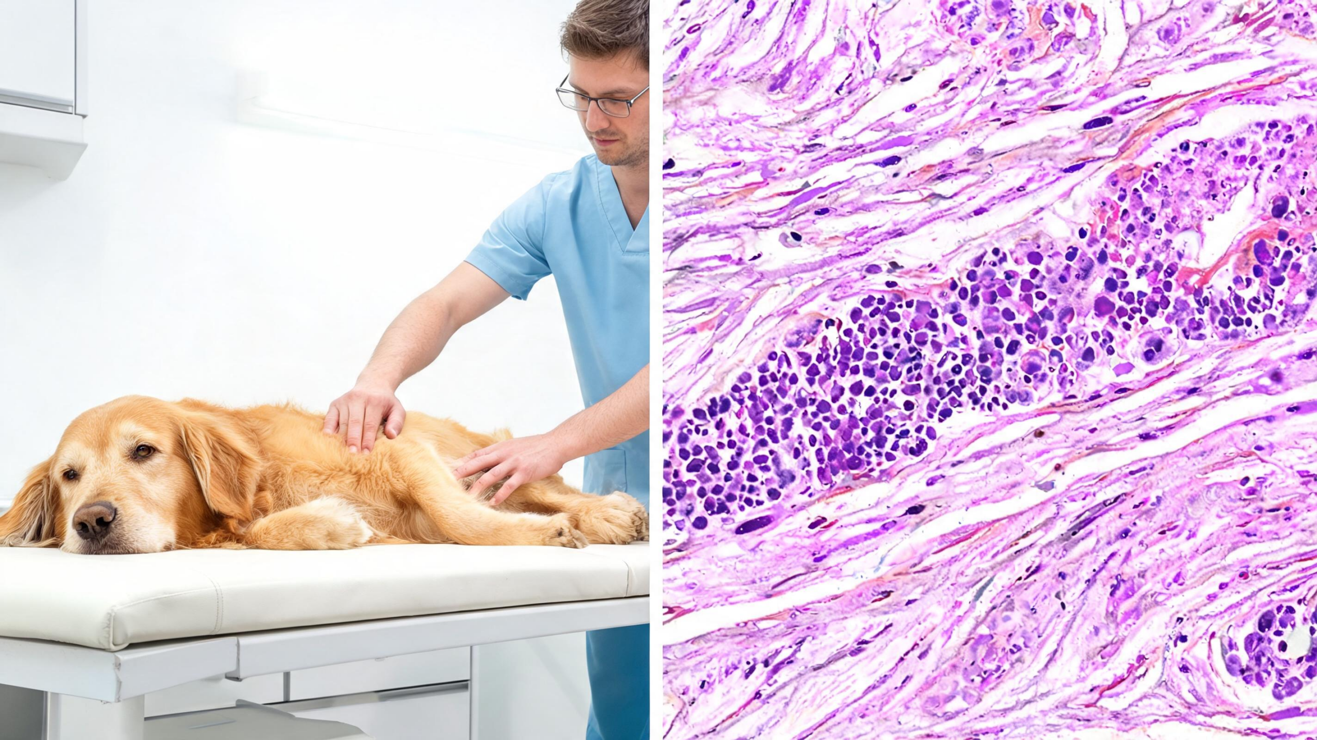
A ten-year-old castrated male mixed breed dog walked into his routine wellness visit with what looked like a minor soft tissue problem: mild swelling around the right hock. Within days, that swelling had exploded into severe pitting edema along an entire hind limb, and within two months the dog was dead. The cause, as veterinarians at Colorado State University’s Veterinary Health System would ultimately determine, was one of the rarest presentations of cancer seen in either veterinary or human medicine: a T-cell lymphoma that had spread extensively through skeletal muscle and that carried an unusual molecular signature, aberrant expression of the Mum-1 protein. The case, published as an open-access report in the journal Veterinary Oncology, offers one of the most detailed clinical accounts yet of a disease so uncommon that only a handful of confirmed cases have ever been documented in dogs.
Primary intramuscular lymphoma, meaning lymphoma that arises in or overwhelmingly involves skeletal muscle rather than lymph nodes or organs, is exceptionally rare across species. In humans, it accounts for less than 1.5 percent of extranodal lymphoma cases, with diffuse large B-cell lymphoma the most common form reported in muscle. Patients typically present with limb swelling, muscle pain, and rapidly worsening symptoms, and tumors have been described in muscles ranging from the sartorius and psoas to the gastrocnemius and soleus. In dogs, the literature is even thinner. Prior to this report, the authors could identify only eight cases of lymphoma with muscular involvement, and two of those lacked a definitive diagnosis. Strikingly, five of the confirmed canine cases were T-cell lymphomas, whereas B-cell disease dominates in human intramuscular cases, hinting at a genuine species difference in the biology of this rare tumor.
The Colorado State patient’s clinical course began deceptively. At the wellness visit, the focal tarsal edema was attributed to a soft tissue injury, and an ulcerated skin lesion on the right lateral neck yielded non-diagnostic cytology. Within hours, the dog was at an emergency clinic, where a 4 by 6 centimeter mass was found in the right inguinal region. Blood work, a chemistry profile, and a tick-borne disease panel were unremarkable. Three days later, diarrhea developed, the inguinal mass had grown to 7 centimeters, and the dog was started on metronidazole and anti-inflammatory prednisone. When he arrived at the oncology service, his right pelvic limb and inguinal region were severely edematous, the mass itself hidden beneath the swelling, and his peripheral lymph nodes were normal in size and texture, an important clue that this was not ordinary nodal lymphoma.
Computed tomography of the limb, abdomen, and thorax revealed the true extent of the disease. Multifocal, isoattenuating masses that enhanced strongly with contrast were scattered diffusely throughout the musculature of the body, involving the semimembranosus, adductor magnus, vastus lateralis, gluteal, gracilis, biceps femoris, epaxial, subscapularis, infraspinatus, and triceps muscles, with individual lesions ranging from 1 to 12 centimeters. Additional small masses sat near the caudal vena cava and adjacent to the ventricular free walls of the heart, though these were deemed too risky to sample. Angiography ruled out a thrombus as the cause of the edema. Ultrasound-guided fine needle aspirates of the thigh masses showed a population of individualized round cells with vacuolated cytoplasm and mild to moderate atypia, suggestive of a discrete cell neoplasm but not definitive. The differential list spanned lymphoma, embryonal rhabdomyosarcoma, extraosseous osteosarcoma, histiocytic sarcoma, and hemangiosarcoma.
Facing rapidly progressive edema, the team biopsied the left thigh masses with tru-cut needles and, before histopathology returned, began empirical chemotherapy with L-asparaginase and doxorubicin, chosen to cover both lymphoma and sarcoma differentials. The response was dramatic: the owners reported improvement within 24 hours and near-complete resolution of the edema within 72 hours. Histopathology, however, was perplexing. Sheets of neoplastic round cells infiltrated between myocytes, frequently isolating individual muscle fibers, with 25 mitoses counted in just two high-powered fields. Immunohistochemistry deepened the mystery. The cells were negative for CD3, CD20, Pax5, Desmin, CD204, and Iba1, but diffusely positive for vimentin, and roughly 75 percent showed strong nuclear reactivity for Mum-1, a marker typically associated with plasma cell tumors and certain B-cell lymphomas.
The decisive tool was PARR, the polymerase chain reaction assay for antigen receptor rearrangements, which detects clonal immune cell populations by examining gene rearrangements. The assay revealed polyclonal immunoglobulin genes but a clonal T-cell receptor arrangement, directly contradicting the plasma cell tumor interpretation suggested by the Mum-1 staining. Additional immunohistochemistry for CD34 and CD30 came back negative, excluding myeloid neoplasms and anaplastic large cell lymphoma, both of which can express Mum-1. The final diagnosis was T-cell lymphoma with aberrant Mum-1 expression and loss of the pan-T-cell marker CD3. Aberrant Mum-1 expression has been described before in canine and feline T-cell lymphoma, and loss of CD3 occurs in roughly 22.8 percent of canine T-cell lymphomas assessed by flow cytometry, but the combination made this case diagnostically treacherous. In total, achieving a definitive diagnosis took 31 days from the first chemotherapy dose.
Treatment continued with single-agent doxorubicin every three weeks, but eleven days after the third dose the disease declared its aggression. A new 5.8 by 5.2 centimeter fixed mass appeared at the base of the left ear with facial swelling, concerning for progression and possible central nervous system involvement, while peripheral lymph nodes remained unaffected. Cytology confirmed the same neoplastic population. The protocol was switched to a combination of lomustine and L-asparaginase, and within two days the facial mass had nearly resolved, only to recur and progress within eight days, eventually surrounding the left eye. A palliative single 6 Gy fraction of radiation therapy was delivered to the left side of the head, followed by one cycle of an MVPP protocol combining mechlorethamine, vinblastine, procarbazine, and prednisolone. Radiation rapidly reduced the facial swelling, and the report notes this is the only documented use of radiation therapy for intramuscular lymphoma in a dog.
The reprieve was brief. One week after radiation and MVPP, the facial swelling had improved and ventral edema was stable, but eleven days later edema progressed along the limbs and ventrum, and for the first time the dog had trouble rising and appeared uncomfortable. His owners elected humane euthanasia 63 days after initial presentation. No necropsy was performed, leaving open the question of whether the intramuscular masses represented a true primary muscular lymphoma or systemic spread, though the authors argue the overwhelming muscle burden, mild lymphadenomegaly, and complete absence of peripheral lymphadenopathy or organomegaly throughout the course favor a primary muscular process. Even at 63 days, the survival represents one of the longest reported for an dog with an aggressive lymphoma and significant intramuscular involvement.
The case carries several lessons for veterinary oncology, and echoes for comparative medicine. Intramuscular lymphoma should be on the differential list for any animal presenting with acutely progressive limb edema and multifocal muscle masses, even when lymph nodes are normal. Diagnosis demands a multimodal arsenal: contrast-enhanced CT to map disease extent, cytology and histopathology to characterize the cells, immunohistochemistry to define the lineage, and PARR or flow cytometry to resolve clonality when marker expression is misleading. The authors acknowledge that, in hindsight, a multiagent protocol such as CHOP, or an alkylator-rich LOPP regimen given the T-cell phenotype, would likely have been the preferred first-line approach had the diagnosis been known earlier. Instead, the urgency of the edema forced empirical monotherapy before the diagnosis was secure, a common dilemma with this tumor type.
Ultimately, the report underscores both the promise and the limits of modern veterinary cancer care. This tumor responded, at least transiently, to L-asparaginase, doxorubicin, lomustine with L-asparaginase, MVPP chemotherapy, and focal radiation, yet every response was measured in days to weeks, and the prognosis remains guarded. With fewer than ten documented canine cases and only a scattering of human reports, no consensus exists on optimal diagnosis or treatment, and each carefully documented case materially expands the field. The Colorado State team hopes that complete reporting of future cases, including imaging, immunophenotyping, clonality testing, and outcomes, will eventually yield a shared framework for this rare and relentless disease, one that in dogs appears to favor T-cells, strike through muscle, and progress faster than clinicians can comfortably respond.
Subject of Research: A rare canine T-cell lymphoma with extensive intramuscular involvement and aberrant Mum-1 expression
Article Title: T-cell lymphoma with significant intramuscular involvement with Mum-1 expression in a dog
Article References: Gutwillig, M., Schaffer, P., Yoon, P., & Vickery, K. (2026). T-cell lymphoma with significant intramuscular involvement with Mum-1 expression in a dog. Veterinary Oncology, 3(1), Article 21. https://doi.org/10.1186/s44356-026-00075-2
Image Credits: AI Generated
DOI: 10.1186/s44356-026-00075-2
Keywords: canine lymphoma, T-cell lymphoma, intramuscular lymphoma, Mum-1, veterinary oncology, immunohistochemistry, PARR, doxorubicin, L-asparaginase, radiation therapy, computed tomography, extranodal lymphoma
Cite Scienmag News
Nathaniel Bowman. (September 10, 2026). Rare Muscle-Infiltrating T-Cell Lymphoma in a Dog Challenges Diagnosis. Scienmag. https://scienmag.com/rare-muscle-infiltrating-t-cell-lymphoma-in-a-dog-challenges-diagnosis/
Nathaniel Bowman. "Rare Muscle-Infiltrating T-Cell Lymphoma in a Dog Challenges Diagnosis." Scienmag, 10 September 2026, https://scienmag.com/rare-muscle-infiltrating-t-cell-lymphoma-in-a-dog-challenges-diagnosis/. Accessed 10 September 2026.
Nathaniel Bowman. "Rare Muscle-Infiltrating T-Cell Lymphoma in a Dog Challenges Diagnosis." Scienmag. September 10, 2026. https://scienmag.com/rare-muscle-infiltrating-t-cell-lymphoma-in-a-dog-challenges-diagnosis/


