


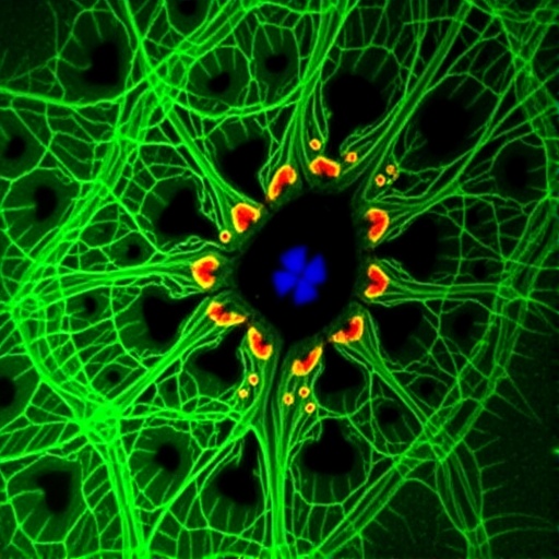
In a development that could reshape how scientists understand and ultimately treat one of the most devastating childhood neurodegenerative conditions, researchers in Norway have created the first human stem cell model of CLN3 Batten disease using astrocytes, the brain’s most abundant glial cells, and uncovered a surprising set of molecular disruptions that place mitochondria, not lysosomes, at the center of the earliest disease changes. The study, published in the Journal of Biomedical Science, was led by Mingyi Yang, Wei Wang and senior authors Magnar Bjørås and Mirta Mittelstedt Leal de Sousa, working across Oslo University Hospital and the Norwegian University of Science and Technology in Trondheim.
CLN3 Batten disease, formally known as juvenile neuronal ceroid lipofuscinosis, is the most common form of a family of inherited lysosomal storage disorders and affects roughly one in 100,000 live births worldwide. It is caused by mutations in the CLN3 gene, with approximately 85 percent of patients carrying the same genetic defect: a deletion of about one kilobase of DNA that removes exons 7 and 8. Children with the disease typically develop normally until between the ages of four and twelve, when vision loss begins. That vision loss progresses to blindness, followed by cognitive decline, motor deterioration, seizures and brain atrophy, culminating in premature death between the ages of 15 and 30. At the cellular level, the disease is marked by the buildup of autofluorescent storage material inside cells, whose major protein component is subunit C of the mitochondrial ATP synthase enzyme, a molecular fingerprint of failing cellular housekeeping.
The CLN3 protein itself is a transmembrane protein that resides primarily in endosomes and lysosomes, the cell’s recycling and degradation compartments, and although it has been linked to cellular homeostasis and neuronal survival, its precise function has remained elusive. For decades, research into the disease has been dominated by a neuron-centric view, focused on the nerve cells that die as the disease progresses. But an accumulating body of evidence has challenged that framing. Studies in mouse models have shown that glial activation precedes the loss of neurons, and that the locations where glial cells become activated accurately predict where neurons will subsequently die. Even more strikingly, laboratory co-culture experiments have demonstrated that glial cells lacking functional CLN3 are toxic to both healthy and mutant neurons, while adding healthy glial cells to mutant neurons largely rescues their survival. In other words, the supportive cells of the brain may be active participants in the neurodegeneration rather than innocent bystanders.
To interrogate the role of astrocytes specifically, the team took skin biopsy fibroblasts from a Norwegian CLN3 patient homozygous for the canonical 1 kb deletion and reprogrammed them into induced pluripotent stem cells using a Sendai virus-based reprogramming kit. Those pluripotent cells were then guided through a carefully choreographed differentiation pipeline: first into neural stem cells using small molecule inhibitors including CHIR99021 and SB431542, then into glial progenitor cells nourished with FGF-2 and EGF, and finally into mature astrocytes driven by leukemia inhibitory factor, EGF and ciliary neurotrophic factor over a maturation period of four weeks. The resulting cells expressed a suite of astrocyte markers — GFAP, S100β, ALDH1L1 and the glutamate transporter EAAT1/GLAST — at levels confirming successful and pure differentiation, while neuronal and oligodendrocyte markers remained minimal. Quantitative PCR confirmed the patient cells carried two mutated CLN3 alleles, and targeted mass spectrometry revealed that the truncated mutant CLN3 protein was not detectable at all in patient-derived cells, confirming that the deletion effectively abolishes CLN3 protein production.
With a validated cellular model in hand, the researchers performed an integrated analysis of both the transcriptome, using whole-transcriptome RNA sequencing, and the proteome, using label-free quantitative mass spectrometry on a timsTOF Pro 2 instrument in PASEF mode. The scale of the effort was considerable: multiple clones from two healthy control individuals and from the patient, each differentiated in triplicate, generated thousands of differentially expressed genes and proteins that could be compared across the developmental trajectory from stem cell to astrocyte. Gene set enrichment analysis of these datasets revealed the study’s central surprise. While conventional wisdom held that CLN3 loss should primarily disrupt lysosomal function, the young astrocytes showed only subtle lysosomal alterations. The lysosomal marker LAMP1 was present at similar levels in patient and control cells, and processes related to lysosomal protein catabolism were enriched to a similar degree in both genotypes during differentiation.
The mitochondrial story was entirely different, and it unfolded in two distinct phases. In the patient’s induced pluripotent stem cells, gene expression data showed underrepresentation of the mitochondrial respiratory chain complex I, accompanied by signatures of negative regulation of TORC1, a master growth and metabolism regulator — a combination previously observed in yeast models lacking the CLN3 homologue. But when the cells matured into astrocytes, the pattern inverted dramatically: proteins belonging to respiratory chain complexes I and IV, including NADH dehydrogenase components and the mitochondrial respirasome, were significantly overrepresented in the patient-derived cells. This contrasts sharply with the downregulation of these same complexes typically observed in CLN3-deficient neurons. Mitochondrial DNA copy number measurements confirmed that the patient astrocytes did not simply contain more mitochondria; instead, they appeared to pack more respiratory supercomplex proteins into their inner mitochondrial membranes, which themselves showed signs of disorganized structure, with genes governing mitochondrial fusion and inner membrane organization downregulated.
This mitochondrial upheaval rippled into lipid metabolism. The team found elevated levels of ELOVL1, the enzyme responsible for elongating very-long-chain saturated fatty acids, suggesting a shift toward production of longer saturated lipid species — a potentially dangerous change, given that elevated ELOVL1 activity has been linked to neurotoxicity in reactive astrocytes through saturated lipid production. At the same time, ELOVL5 levels dropped slightly, implying reduced synthesis of polyunsaturated fatty acids that maintain membrane fluidity. Fatty acid synthesis enzymes such as ACACA and FASN were downregulated, while the β-oxidation enzymes ACOX1 and ACADVL were upregulated, pointing to a metabolic pivot away from lipid construction and toward fatty acid burning. Notably, ACADS, which handles short-chain fatty acid oxidation and is normally upregulated during healthy astrocyte differentiation, failed to rise in the patient cells, suggesting the metabolic adaptation itself was broken. Because astrocytes account for the oxidation of free fatty acids that represents roughly 20 percent of the brain’s total energy expenditure, these defects strike at the heart of cerebral energy economics.
The dysregulated fat metabolism and aberrant respiratory chain activity came with a third signature: an intensified oxidative stress response. Levels of glutathione synthetase, which produces the cell’s principal antioxidant, were substantially increased in patient cells, as were the glutathione-conjugating enzymes GSTK1 and GSTZ1, along with catalase, NQO1 and the mitochondrial superoxide dismutase SOD2. The researchers interpret this as a compensatory counterattack against elevated reactive oxygen species generated by runaway peroxisomal β-oxidation — ACOX1 activity produces hydrogen peroxide as a byproduct — combined with impaired detoxification of partially oxidized fatty acid intermediates. Prior studies have documented increased oxidative stress in CLN3 lymphoblasts and fibroblasts, and work in fruit flies has shown that CLN3 loss increases vulnerability to oxidative damage while CLN3 overexpression confers resistance, lending independent support to the connection the Norwegian team has now drawn in human astrocytes.
Beyond metabolism, the analysis surfaced evidence of deeper epigenetic rewiring. A multifactorial statistical analysis disentangling the effects of genotype and cell type identified 363 genes whose expression patterns diverge specifically in the patient during astrocyte differentiation, with significant overrepresentation of genes involved in histone H3 and H4 lysine methylation. In patient cells, chromatin-related terms associated with open, transcriptionally active states — such as binding of acetylated histones — were underrepresented, while repressive machinery, including polycomb group complexes and heterochromatin-associated terms, was upregulated. The authors suggest this reflects a global shift toward a more compacted, transcriptionally restrictive chromatin landscape, potentially locking developing astrocytes into aberrant functional states. Additional dysregulation was seen in extracellular matrix genes involved in remodeling, synaptic support and neuroinflammatory signaling, hinting that mutant astrocytes undergo a reactive transformation that could compromise their support of neurons even before overt cell death begins.
The study is not without limitations, which the authors acknowledge candidly. All patient-derived material came from a single individual, so some observed phenotypes could reflect that patient’s unique genetic background rather than CLN3 loss alone, and extending the work to additional patient lines or gene-corrected isogenic controls would strengthen the conclusions. Nevertheless, the findings carry significant therapeutic implications. If mitochondrial dysfunction in astrocytes indeed precedes the lysosomal breakdown that defines advanced disease, then interventions aimed at restoring mitochondrial health — improving respiration, normalizing lipid metabolism, or bolstering antioxidant capacity — could preserve astrocyte function, and through it neuronal survival, potentially delaying disease progression. The team proposes that future work in more complex human models, such as brain organoids incorporating multiple cell types, should be used to establish the precise temporal relationship between astrocytic mitochondrial failure and neuronal death. Because mitochondrial dysfunction is a common thread linking Alzheimer’s, Parkinson’s, Huntington’s and ALS, insights from this rare pediatric disease may ultimately illuminate mechanisms of far more common neurodegenerative conditions, including those associated with aging, underscoring once again that understanding rare diseases can pay dividends well beyond the patient communities they directly affect.
Cite Scienmag News
Drew Townsend. (September 9, 2026). Astrocyte model of Batten disease reveals mitochondrial and metabolic defects. Scienmag. https://scienmag.com/astrocyte-model-of-batten-disease-reveals-mitochondrial-and-metabolic-defects/
Drew Townsend. "Astrocyte model of Batten disease reveals mitochondrial and metabolic defects." Scienmag, 9 September 2026, https://scienmag.com/astrocyte-model-of-batten-disease-reveals-mitochondrial-and-metabolic-defects/. Accessed 9 September 2026.
Drew Townsend. "Astrocyte model of Batten disease reveals mitochondrial and metabolic defects." Scienmag. September 9, 2026. https://scienmag.com/astrocyte-model-of-batten-disease-reveals-mitochondrial-and-metabolic-defects/


