


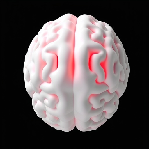
Pediatric head computed tomography has become an indispensable tool in emergency medicine, offering rapid diagnosis in cases of trauma, hydrocephalus, seizures, and suspected intracranial pathology. Yet the very technology that makes urgent neuroimaging possible also exposes children to ionizing radiation at doses that carry disproportionately greater risk than in adults, because developing tissues are markedly more radiosensitive and because children have longer lifespans over which stochastic radiation effects may manifest. This tension between diagnostic benefit and long-term risk has driven radiologists and medical physicists to seek ever-finer methods of protocol optimization, dose reduction, and image quality validation. A persistent obstacle, however, has been the lack of test objects that faithfully reproduce pediatric anatomy and attenuation. Most commercially available phantoms are modeled on adult bodies, use uniform acrylic cylinders, or approximate children by simply scaling down adult geometries, none of which captures the true shape, size, and radiological behavior of a child’s brain. A team of researchers in Morocco has now addressed this gap with a rigorously validated, three-dimensional, patient-specific pediatric brain phantom built directly from clinical CT data, described in a new study published in Pediatric Radiology.
The research, led by Hamza Sekkat of Université Hassan 1er in Settat and colleagues, began with a fully anonymized head CT examination of a 5-year-old child, archived from routine clinical practice. Using the open-source medical image computing platform 3D Slicer, the team performed a careful segmentation of the brain parenchyma, isolating the brain tissue from the skull, cerebrospinal fluid spaces, and surrounding structures. The resulting segmented volume was exported as a standard tessellation language, or STL, mesh — the file format universally used in additive manufacturing — and digitally partitioned into two cerebral hemispheres. This hemispheric split was a deliberate design decision: it allows the phantom to be cast in parts and later assembled, giving researchers access to internal surfaces and making future insertion of simulated lesions, ventricles, or other structures far more practical than a single monolithic cast would allow.
From the STL files, the team three-dimensional-printed a polylactic acid, or PLA, master model of each hemisphere. These prints served as the positive forms around which a reinforced silicone mold was fabricated. Silicone was chosen for its flexibility and dimensional stability, allowing the delicate contours of cortical gyri and the interhemispheric fissure to be captured faithfully while still permitting clean demolding of the final cast. This two-stage approach — print a master, mold the negative, cast the surrogate — is a well-established strategy in anthropomorphic phantom fabrication, but its application to a genuinely patient-specific pediatric brain, rather than a generic or adult-derived template, is what distinguishes the present work. The method is also low-cost and reproducible, relying on widely available desktop printing and casting materials rather than specialized industrial equipment.
The central materials-science challenge was to formulate a brain-mimicking substance whose X-ray attenuation matches that of living brain parenchyma across clinically relevant energies. The researchers selected an epoxy resin system as the base matrix, prized in tissue-equivalent materials for its homogeneity, dimensional stability, and tunable chemistry. To bring the attenuation down to soft-tissue levels — pure epoxy is typically denser and more attenuating than brain tissue — they modified the resin with acetone. The addition of a volatile organic solvent to an epoxy system is a known technique for lowering effective density, but it must be handled with care: residual solvent can affect cure kinetics, porosity, and long-term stability of the cast material. The team’s formulation produced a homogeneous solid surrogate that poured and cured within the silicone molds without significant voids or segregation, yielding casts with the fine surface detail of the original segmented anatomy.
When the assembled hemispheres were scanned and compared with the source patient’s images, the geometric fidelity was striking. The fabricated phantom retained 96.85 percent of the original patient-derived brain volume — 1,129.15 cubic centimeters in the phantom versus 1,165.89 cubic centimeters in the segmented clinical dataset. The main bilateral morphology, including the gross symmetry of the hemispheres and the external contours, was preserved. For dosimetry and protocol-optimization studies, this kind of volumetric and morphological accuracy matters enormously: scattering conditions, beam hardening, and noise texture in reconstructed images all depend on the size and shape of the attenuating object, and a phantom that is even modestly mis-sized can mislead dose estimates and image quality benchmarks.
Attenuation matching was evaluated across a deliberately broad range of acquisition conditions. The phantom was scanned at four tube voltages spanning standard pediatric practice — 80, 100, 120, and 140 kilovolt peak, or kVp. Region-of-interest measurements were placed across both hemispheres to quantify CT numbers, the Hounsfield unit values that encode local X-ray attenuation. At 120 kVp, the voltage most commonly used in pediatric head protocols and the voltage of the original source scan, the phantom’s mean CT number was 44.94 ± 1.38 HU, compared with 40.66 ± 4.13 HU in the source pediatric brain parenchyma — an absolute difference of just 4.28 HU, well within the tolerance generally considered acceptable for soft-tissue equivalence. Across the full acquisition series, mean CT numbers ranged from 18.55 ± 1.14 HU at 80 kVp to 51.04 ± 0.93 HU at 140 kVp, and the CT-derived mass attenuation coefficients decreased from 0.1664 ± 0.0002 to 0.1445 ± 0.0001 square centimeters per gram over the 80-to-140 kVp range. This systematic energy dependence is precisely the kind of data needed to validate the phantom not just at one nominal setting but across the entire span of voltages a pediatric department might deploy.
To push the validation beyond the scanner itself, the team turned to computational physics. They used the Particle and Heavy Ion Transport code System — PHITS, a widely respected Monte Carlo radiation transport code — to simulate photon transmission through the surrogate material with a mono-energetic transmission method covering photon energies from 15 to 150 kiloelectronvolts, a range that envelops the effective spectra of all diagnostic X-ray tubes. The simulated attenuation coefficients were then benchmarked against two independent theoretical references: the PhyX–Photon Shielding and Dosimetry, or PhyX-PSD, online platform, and the National Institute of Standards and Technology’s XCOM database, the canonical repository of photon cross sections. Across the entire predefined 15-to-150 keV diagnostic energy window, the PHITS-derived values agreed with the theoretical references to within ±5 percent relative difference. This triple agreement — physical CT measurements on a real scanner, Monte Carlo simulation, and tabulated reference data — constitutes a physics-validated characterization of the surrogate’s energy-dependent attenuation, giving researchers confidence that the phantom will behave like real brain tissue under beam conditions it has never physically been exposed to.
The implications for pediatric imaging research are substantial. With a validated, anatomically faithful, attenuationally accurate pediatric brain phantom, departments can benchmark low-dose protocols, iterative reconstruction and deep-learning reconstruction algorithms, dual-energy and photon-counting techniques, and organ-dose estimation methods under realistic pediatric conditions rather than adult approximations. It also opens the door to radiation therapy applications, where patient-specific phantoms are used to verify treatment planning calculations, and to training, where residents and technologists can practice scanning and protocol selection on a realistic child-sized head without exposing any patient. The authors frame the present brain component as the first module of a larger program: the same CT-derived, mold-and-cast workflow will be extended to the pediatric skull — building on the group’s earlier work on cranial bone substitutes — and to other head structures, ultimately yielding a modular, complete-head phantom in which each tissue component has been independently validated.
The study also demonstrates a workflow that any reasonably equipped medical physics laboratory could adopt. The entire pipeline — anonymized CT acquisition, open-source segmentation in 3D Slicer, STL export, desktop 3D printing of a PLA master, silicone molding, epoxy-acetone casting, and Monte Carlo verification with freely available tools — avoids proprietary software and expensive commercial phantom suppliers. In settings where pediatric imaging research is under-resourced, including much of the developing world, this accessibility could be transformative. The work was supported by the National Center for Scientific and Technical Research in Morocco through its PhD-Associate Scholarship program, and the authors report no competing interests. The study used exclusively anonymized retrospective imaging data, posing no additional risk to any patient.
What emerges from this research is both a concrete artifact and a methodological template: proof that a child’s brain can be faithfully reproduced in a test object that matches its shape to within a few percent of its volume and its CT attenuation to within a few Hounsfield units at matched energy, with physics-validated behavior across the full diagnostic photon range. As dose-reduction technologies such as photon-counting detectors and artificial-intelligence reconstruction continue to advance, the ability to test them against true pediatric anatomy — rather than scaled-down adult surrogates — will be essential to ensuring that the smallest and most vulnerable patients receive the safest possible imaging. The Moroccan team’s phantom is a quiet but consequential step toward that standard, and its modular design promises a complete pediatric head phantom in which every tissue equivalent is held to the same rigorous, multi-validated benchmark.
Cite Scienmag News
Colin Clarke. (September 5, 2026). New 3D pediatric brain phantom validated for CT neuroimaging research. Scienmag. https://scienmag.com/new-3d-pediatric-brain-phantom-validated-for-ct-neuroimaging-research/
Colin Clarke. "New 3D pediatric brain phantom validated for CT neuroimaging research." Scienmag, 5 September 2026, https://scienmag.com/new-3d-pediatric-brain-phantom-validated-for-ct-neuroimaging-research/. Accessed 5 September 2026.
Colin Clarke. "New 3D pediatric brain phantom validated for CT neuroimaging research." Scienmag. September 5, 2026. https://scienmag.com/new-3d-pediatric-brain-phantom-validated-for-ct-neuroimaging-research/


