


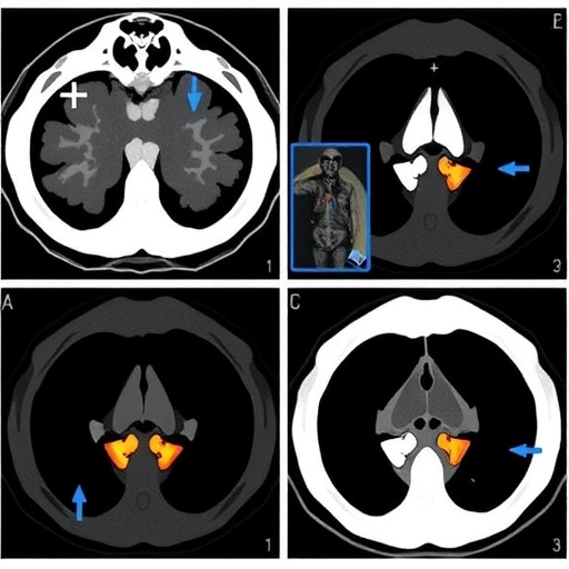
Multiple myeloma, a cancer of antibody-producing plasma cells, has long challenged physicians because it behaves less like a single disease than a shifting ecosystem of genetically distinct tumor populations. Some patients respond durably to treatment, while others progress rapidly despite apparently similar laboratory results and imaging findings. A new study published in the European Journal of Nuclear Medicine and Molecular Imaging reports a potential way to make one of the disease’s most important imaging measurements faster, more reproducible, and easier to combine with genomic information. Researchers from the University of Bologna and affiliated institutions developed an artificial-intelligence-assisted pipeline that automatically calculates Deauville Scores from baseline [¹⁸F]FDG PET/CT scans and uses them, together with blood tests and chromosomal abnormalities, to predict progression-free survival.
The Deauville Score is a five-point scale originally developed for interpreting fluorodeoxyglucose PET scans in lymphoma. It compares the metabolic activity of suspected disease with normal reference tissues, especially the mediastinal blood pool and liver. In multiple myeloma, the same principle can be applied to different patterns of disease, including diffuse bone-marrow infiltration, focal bone lesions, paramedullary growth extending beyond bone, and extramedullary disease in soft tissues. A score of 1 indicates no abnormal uptake, while scores of 4 or 5 indicate activity clearly exceeding normal reference uptake. Although clinically useful, Deauville assessment is usually based on visual interpretation, leaving room for differences between readers, institutions, scanners, and reporting habits. The new system, named autoDS-PET, is designed to turn that visual framework into a standardized computational measurement.
The investigation included 165 patients with newly diagnosed multiple myeloma who underwent FDG PET/CT before starting treatment. All participants also had copy number alteration testing and routine blood analyses. The cohort was assembled retrospectively from patients diagnosed at a Bologna medical center between 2007 and 2021, spanning four different PET/CT scanner models and a long period of changing treatment strategies. The researchers analyzed five anatomical compartments: vertebral bone marrow, marrow in the long bones, focal lesions, paramedullary lesions, and extramedullary lesions. The pipeline operated fully automatically for vertebral and long-bone marrow, while lesion-based analysis remained semi-automatic because a nuclear medicine physician manually outlined the focal, paramedullary, and extramedullary lesions requiring evaluation.
Behind the scenes, the software performs a sequence of image-processing steps that would normally demand considerable technical labor. DICOM imaging files are converted into NIfTI format, PET data are normalized to body-weight-based standardized uptake values, and PET and CT images are rigidly registered so that corresponding anatomical structures align precisely. The system then uses TotalSegmentator, a deep-learning model based on the nnU-Net architecture, to identify vertebrae, vertebral bodies, the aorta, liver, femurs, and humeri on CT. These automatically generated masks are refined with mathematical morphology carried out in physical space, rather than simply by counting voxels. That distinction matters because medical images can have unequal voxel dimensions, and a three-voxel erosion does not represent the same physical distance in every scan.
For vertebral marrow, the program combines the L3, L4, and L5 vertebrae, isolates the vertebral bodies, excludes posterior elements, and erodes the resulting mask by 3 millimeters to reduce contamination from cortical bone and partial-volume effects. The mediastinal blood-pool reference is obtained from the aorta within the T4-to-T8 vertebral region, with heart tissue removed to limit spill-in from cardiac activity and a 4-millimeter erosion used to focus on the vessel lumen. The liver mask is cleaned, filled, and eroded by 10 millimeters to provide a stable parenchymal reference. For long bones, the system crops the femur and humerus masks to their central 60 percent, approximating the diaphysis, then removes the cortical shell with additional erosion. The resulting region captures appendicular marrow that is often overlooked in conventional myeloma assessment.
The software also makes a deliberate distinction between diffuse marrow disease and discrete lesions. For vertebral and long-bone marrow, it uses the 95th percentile of SUV rather than the single hottest voxel, limiting the influence of isolated outliers or image noise. For focal, paramedullary, and extramedullary lesions, it uses maximum SUV because peak metabolic activity may be clinically meaningful in a compact lesion. Median uptake within the mediastinal blood pool and liver provides the reference values. The program then assigns the corresponding Deauville category automatically. Importantly, the investigators report that the pipeline completed successfully in all 165 patients without manual intervention for the fully automated components, suggesting that the workflow is technically feasible even across heterogeneous clinical imaging data.
The prognostic results revealed that the location of metabolically active disease may matter as much as its intensity. High Deauville Scores in long-bone marrow, paramedullary lesions, and extramedullary lesions were associated with shorter progression-free survival in univariate analyses. Long-bone involvement was particularly notable: although 86.1 percent of patients fell into the low-score group, those with scores of 4 or 5 had significantly worse outcomes, with a log-rank p-value of 0.019. Extramedullary disease was uncommon, present at a high score in only 6.1 percent of patients, but showed the strongest association with poor progression-free survival, with p=0.001. Paramadullary disease was also significant, while the score of the hottest focal lesion alone did not distinguish outcomes. This may indicate that the number and overall burden of focal lesions are more informative than the metabolic intensity of one “hottest” lesion.
The investigators next combined the imaging variables with age, sex, hemoglobin, platelet count, and three recurrent copy number alterations: amplification of chromosome arm 1q, deletion of 13q, and deletion of 17p. They also tested an interaction between amp(1q) and del(13q), which can occur together and may identify biologically aggressive disease. A penalized Cox proportional-hazards model was evaluated using repeated nested cross-validation, a design intended to estimate performance on previously unseen patients while reducing optimism caused by model tuning. The full model achieved an Uno concordance index of 0.710, compared with 0.672 for a reduced model containing only non-imaging variables. Its integrated Brier score was 0.175, indicating better-than-noninformative calibration. In practical terms, the imaging data added measurable prognostic information beyond blood counts and genomic markers, although the performance represents moderate discrimination rather than a definitive clinical prediction system.
In a separate multivariable analysis designed to estimate the contribution of individual features, five factors emerged as independent prognostic variables: age, hemoglobin, vertebral marrow Deauville Score, paramedullary Deauville Score, and amp(1q). Increasing age was associated with higher risk of progression, while higher hemoglobin was protective, consistent with the relationship between anemia, marrow infiltration, and impaired blood production. Higher vertebral marrow and paramedullary scores were also linked to increased risk. Amplification of 1q, detected in 60 patients, carried a hazard ratio of 1.786. The apparently protective hazard ratio for del(17p) was not statistically significant and was interpreted cautiously because only 11 patients carried the abnormality. Similarly, the interaction between amp(1q) and del(13q) did not reach significance, likely reflecting limited statistical power rather than evidence that the combination lacks biological importance.
The study’s authors emphasize that autoDS-PET is not yet a replacement for clinical expertise or a validated treatment-selection device. The cohort came from a single center, the analysis was retrospective, genomic testing covered only three copy number alterations, and the study period included substantial changes in frontline therapy. Lesion detection and segmentation also remain partly dependent on expert input, meaning that full automation has not yet been achieved. Rare extramedullary disease and del(17p) events make some estimates unstable, and the model requires external validation in independent populations using contemporary treatment regimens and standardized acquisition protocols. Nevertheless, the work points toward a compelling direction for precision oncology: rather than treating PET/CT as a collection of subjective images, clinicians could use automated, anatomically specific metabolic scores as structured data. By connecting disease distribution, functional activity, blood-cell production, and tumor genetics, the approach may help transform a routine scan into a reproducible map of myeloma biology. The autoDS-PET code is being released as an open-source research tool, creating an opportunity for other centers to test, challenge, and improve the framework before it can move toward clinical deployment.
Cite Scienmag News
Ophelia Keating. (August 26, 2026). Automated Baseline FDG PET/CT Deauville Scoring Predicts Progression-Free Survival in Multiple Myeloma. Scienmag. https://scienmag.com/automated-baseline-fdg-pet-ct-deauville-scoring-predicts-progression-free-survival-in-multiple-myeloma/
Ophelia Keating. "Automated Baseline FDG PET/CT Deauville Scoring Predicts Progression-Free Survival in Multiple Myeloma." Scienmag, 26 August 2026, https://scienmag.com/automated-baseline-fdg-pet-ct-deauville-scoring-predicts-progression-free-survival-in-multiple-myeloma/. Accessed 4 September 2026.
Ophelia Keating. "Automated Baseline FDG PET/CT Deauville Scoring Predicts Progression-Free Survival in Multiple Myeloma." Scienmag. August 26, 2026. https://scienmag.com/automated-baseline-fdg-pet-ct-deauville-scoring-predicts-progression-free-survival-in-multiple-myeloma/


