


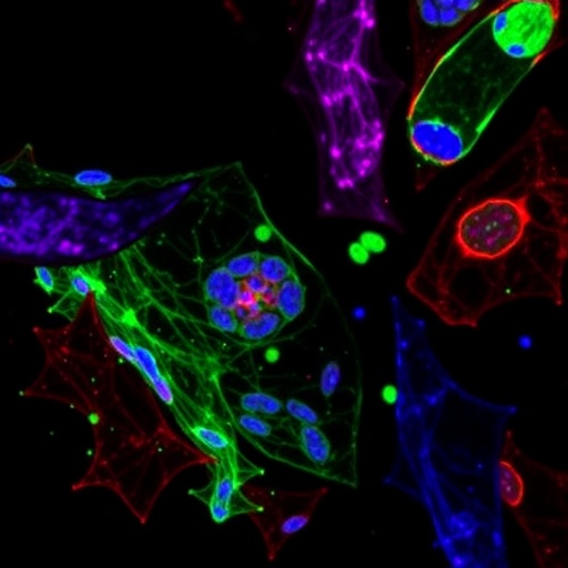
Tumors do not grow in isolation. They develop within a complex ecosystem of blood vessels, connective tissue, signaling molecules and immune cells that can either restrain cancer or help it spread. Among the most influential residents of this ecosystem are tumor-associated macrophages, or TAMs—immune cells recruited into tumors and reshaped by local conditions. A new study published in the Journal of Molecular Medicine reports that a signaling protein called Gsα can determine whether these macrophages behave more like cancer-fighting cells or tumor-supporting cells. According to the researchers, losing Gsα specifically in macrophages accelerated tumor growth and metastasis in mouse models, while the protein promoted an inflammatory, antitumoral macrophage program through the MAPK signaling pathway.
Macrophages are highly adaptable cells. Rather than existing in a single fixed state, they respond continuously to signals from damaged tissue, cancer cells, cytokines, metabolites and neighboring immune cells. In simplified laboratory terminology, macrophages with an M1-like profile are associated with inflammatory and antimicrobial activity, whereas M2-like macrophages are often linked to tissue repair, immune suppression and tumor progression. In real tumors, macrophage states form a spectrum rather than two sharply separated categories. Nevertheless, the balance between inflammatory and immunosuppressive functions can strongly influence the outcome of cancer. TAMs that suppress immune responses may protect malignant cells from attack, stimulate blood-vessel formation and assist invasion into surrounding tissues. Reprogramming these cells has therefore become an important objective in cancer immunotherapy.
The new work focuses on Gsα, the alpha subunit of the stimulatory heterotrimeric G protein. This molecule is best known as a component of signaling downstream from G protein-coupled receptors, a vast family of cell-surface receptors that detect hormones, neurotransmitters, lipids and other extracellular signals. When activated, Gsα commonly stimulates adenylyl cyclase, increasing intracellular cyclic AMP and activating downstream effectors such as protein kinase A. However, G protein signaling is not confined to a single linear route. Depending on the receptor, cellular context and regulatory proteins present, Gsα-associated signals can influence several networks, including the mitogen-activated protein kinase pathway. MAPK signaling includes interconnected kinase cascades such as ERK, p38 and JNK, which regulate gene expression, differentiation, stress responses and inflammatory behavior.
To investigate the role of Gsα in macrophages, the researchers used mice in which the protein was selectively removed from these immune cells. These animals, referred to as GsαMKO mice, were compared with control mice carrying the intact Gsα gene. The team examined tumor development using two widely used experimental systems: B16 melanoma cells and MC38 colorectal cancer cells. In both models, the absence of macrophage Gsα was associated with faster tumor growth. Experiments involving metastatic B16 disease also indicated a greater burden of cancer spread in mice lacking Gsα in macrophages. These findings suggest that the protein affects more than the size of the primary tumor; it may also influence the ability of the tumor microenvironment to support dissemination and colonization of distant organs.
The researchers then examined the molecular identity and behavior of macrophages inside the tumors. Macrophages containing Gsα showed increased expression of CD86, CCR5, Il1b and Nos2, genes and proteins commonly associated with inflammatory activation and immune stimulation. CD86 can provide important co-stimulatory signals during interactions between antigen-presenting cells and T cells. CCR5 is a chemokine receptor involved in immune-cell trafficking, while Il1b encodes interleukin-1 beta, a potent inflammatory mediator. Nos2, also known as inducible nitric oxide synthase, enables macrophages to produce nitric oxide, a reactive molecule involved in antimicrobial and immune effector functions. In contrast, Gsα activity was associated with lower levels of CD206 and Il10, markers linked in this context to alternative, immunosuppressive macrophage behavior.
These changes were not merely molecular labels. The study indicates that Gsα-positive macrophages were better able to contribute to antitumor immunity. Their altered chemokine-receptor profile could affect how macrophages are recruited and positioned within tumors, while their inflammatory gene program could improve their ability to oppose malignant cells. The researchers also observed increased effector activity among CD8-positive T cells in tumors from control animals compared with animals lacking macrophage Gsα. CD8-positive T cells are cytotoxic lymphocytes capable of recognizing and killing abnormal cells, but their effectiveness can be weakened by suppressive conditions within the tumor microenvironment. The results suggest that macrophage Gsα may indirectly strengthen T-cell responses by making the surrounding immune environment less tolerant of cancer.
At the mechanistic level, the investigators linked this macrophage reprogramming to MAPK activity. They reported that Gsα promoted phosphorylation of ERK, p38 and JNK—chemical modifications that activate these kinases and allow them to transmit signals toward the nucleus and other cellular targets. Once activated, MAPK pathways can alter transcription factors and inflammatory gene networks, changing how macrophages respond to tumor-derived signals. The simultaneous involvement of ERK, p38 and JNK is notable because these branches can control overlapping yet distinct aspects of macrophage biology. ERK often participates in proliferation and differentiation signals, p38 is strongly associated with stress and inflammatory responses, and JNK can regulate cytokine production, apoptosis and transcriptional remodeling. Together, their activation may help maintain the proinflammatory state observed in macrophages containing Gsα.
Additional cell-based experiments supported the conclusion that the effect was intrinsic to macrophages rather than simply a consequence of unrelated differences between the animals. Bone marrow-derived macrophages from Gsα-deficient mice displayed altered activation patterns, and restoring exogenous Gsα changed the molecular profile of these cells. The study also used tumor-conditioned media, which contains soluble factors released by cancer cells, to model some of the signals macrophages encounter in the tumor microenvironment. These experiments point to a system in which cancer-derived signals can push macrophages toward tumor-supporting behavior when Gsα is absent, whereas Gsα helps preserve or restore inflammatory functions. The investigators further reported increased CD31 expression in tumors from GsαMKO mice, consistent with enhanced vascular features that could facilitate tumor expansion and metastatic escape, although the precise relationship between macrophage Gsα and blood-vessel formation requires further study.
The findings are especially relevant because many current cancer treatments focus primarily on malignant cells or on immune checkpoints, such as the PD-1 and PD-L1 pathway. Checkpoint inhibitors can release T cells from inhibitory signals, but their success depends on the broader immune environment. Immunosuppressive TAMs are one reason tumors may remain resistant even when cytotoxic lymphocytes are present. A therapy designed to preserve Gsα activity in macrophages, enhance its downstream signaling or selectively activate the relevant MAPK branches could theoretically complement existing immunotherapies. However, the study does not establish a treatment for patients, and directly manipulating G protein signaling would carry substantial risks. Gsα operates in many tissues and participates in physiological processes ranging from hormone responses to metabolism, so a systemic drug could produce effects far beyond the tumor. Any future strategy would need to target macrophages with high precision and determine which receptors or intracellular intermediates connect Gsα to ERK, p38 and JNK in different cancers.
The authors emphasize that their conclusions arise from B16 and MC38 mouse models and from experimental macrophage systems. Human tumors contain diverse macrophage populations shaped by genetics, treatment history, tissue origin and metabolic conditions, and these cells may not respond identically to Gsα manipulation. The study also highlights an important complexity in cell signaling: the same molecular pathway can have different consequences depending on the cell type and biological setting. While Gsα-associated cyclic AMP signaling has been linked in other contexts to anti-inflammatory or M2-like macrophage behavior, this work identifies a macrophage-specific role in which Gsα supports inflammatory antitumor activity through MAPK phosphorylation. Further research will be needed to validate Gsα expression and MAPK activity in human TAMs, establish whether the relationship predicts patient outcomes and determine whether selectively reprogramming this pathway can improve responses to immunotherapy without provoking harmful inflammation. For now, the study adds Gsα to the growing list of molecular switches that may decide whether the immune cells surrounding a tumor become its allies—or its enemies.
Cite Scienmag News
Nathaniel Bowman. (August 26, 2026). Macrophage Gsα Deficiency Accelerates Tumor Progression Through MAPK Signaling. Scienmag. https://scienmag.com/macrophage-gs%ce%b1-deficiency-accelerates-tumor-progression-through-mapk-signaling/
Nathaniel Bowman. "Macrophage Gsα Deficiency Accelerates Tumor Progression Through MAPK Signaling." Scienmag, 26 August 2026, https://scienmag.com/macrophage-gs%ce%b1-deficiency-accelerates-tumor-progression-through-mapk-signaling/. Accessed 4 September 2026.
Nathaniel Bowman. "Macrophage Gsα Deficiency Accelerates Tumor Progression Through MAPK Signaling." Scienmag. August 26, 2026. https://scienmag.com/macrophage-gs%ce%b1-deficiency-accelerates-tumor-progression-through-mapk-signaling/


