


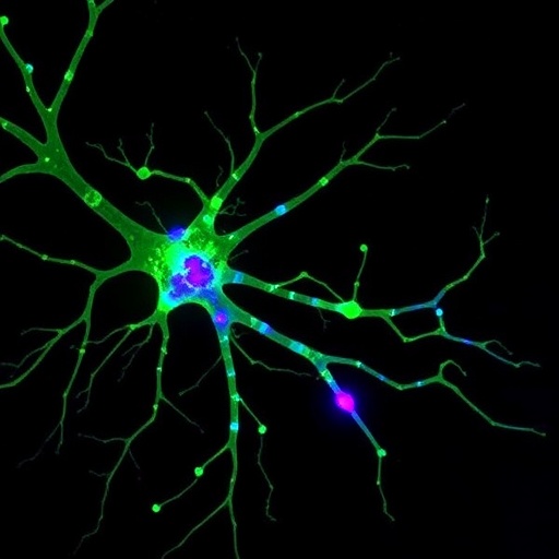
A team led by Del Popolo and colleagues reports that aggregated α-synuclein—an established driver of Parkinson’s disease pathology—does not affect dopamine neurons uniformly. Using acute mouse brain slices, the researchers compared how distinct α-synuclein aggregate preparations reshape the electrical behavior of neurons from two midbrain hubs: the substantia nigra pars compacta (SNpc) and the ventral tegmental area (VTA). The work highlights a cell-type selectivity that may help explain why some dopaminergic circuits degenerate earlier than others.
To probe circuit-specific vulnerability, the investigators performed electrophysiological recordings from dopaminergic neurons in SNpc and VTA within maintained slice preparations. Neurons were challenged with α-synuclein aggregates, and firing patterns were monitored alongside synaptic and membrane properties. This approach allowed the team to distinguish direct effects on intrinsic excitability from changes that could be driven by altered network input.
The study finds that α-synuclein aggregates produce different electrophysiological signatures depending on neuronal location. In SNpc neurons, aggregate exposure shifted firing dynamics in a manner consistent with disrupted voltage-dependent conductances, including changes in action potential timing and spike regularity. These alterations suggest that the aggregates may interfere with the balance of inward and outward ionic currents that normally stabilize pacemaking-like activity.
In contrast, VTA dopaminergic neurons displayed a contrasting response profile. While aggregate treatment still modified electrical activity, the direction and magnitude of the changes differed from SNpc. The authors interpret this divergence as evidence that molecular interactions between aggregates and cell-specific membrane or synaptic machinery are not interchangeable across dopaminergic subtypes.
Beyond firing frequency, the team examined features linked to how neurons respond to stimulation. Aggregate-related effects on excitability appeared to alter how SNpc and VTA neurons integrate inputs and convert them into spiking output. Such differences could translate into circuit-level changes in dopamine release patterns, with potential implications for motor versus reward-related dysfunctions.
The findings also underscore the value of acute slice electrophysiology for separating early, functional consequences of α-synuclein species from slower degenerative processes. Because the experiments are performed shortly after aggregate exposure, the results point to rapid physiological disruption rather than solely chronic neurodegeneration.
The researchers emphasize that not all α-synuclein “aggregate” preparations behave identically. By using aggregate-dependent challenges and comparing two anatomically distinct dopaminergic populations, the study provides a framework for linking aggregate chemistry to specific neuronal phenotypes.
Overall, the work frames α-synuclein as a selective perturbing agent whose physiological impact depends on where the target neuron resides in the midbrain. If replicated and extended in vivo, these circuit-selective effects could inform why Parkinson’s symptoms emerge with particular spatial and functional patterns—and how future therapeutics might be tuned accordingly.
Cite Scienmag News
Cassandra Pierce. (July 28, 2026). Alpha-synuclein Aggregates Differentially Alter Electrophysiology of SNpc and VTA Neurons. Scienmag. https://scienmag.com/alpha-synuclein-aggregates-differentially-alter-electrophysiology-of-snpc-and-vta-neurons/
Cassandra Pierce. "Alpha-synuclein Aggregates Differentially Alter Electrophysiology of SNpc and VTA Neurons." Scienmag, 28 July 2026, https://scienmag.com/alpha-synuclein-aggregates-differentially-alter-electrophysiology-of-snpc-and-vta-neurons/. Accessed 7 September 2026.
Cassandra Pierce. "Alpha-synuclein Aggregates Differentially Alter Electrophysiology of SNpc and VTA Neurons." Scienmag. July 28, 2026. https://scienmag.com/alpha-synuclein-aggregates-differentially-alter-electrophysiology-of-snpc-and-vta-neurons/


