


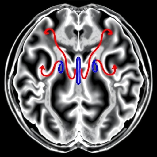
Microgravity during spaceflight can trigger headward fluid shifts and disrupt cerebral blood flow, potentially contributing to visual impairment, spatial disorientation, and cognitive changes in astronauts. Yet, many studies have focused on later stages of simulated microgravity, leaving the early “postural transition” period less understood—especially how blood-flow fluctuations link to changes in upstream arteries.
A team from Capital Medical University, collaborating with the National Space Science Center, addressed this gap in a study published in Space: Science & Technology. Using a head-down bed rest (−6°) protocol, they examined early cerebrovascular coupling mechanisms in 33 healthy male volunteers. Measurements were taken at baseline and after 12 hours to capture rapid hemodynamic adaptation.
To overcome constraints of spaceflight research, the investigators combined Doppler ultrasound with three-dimensional pseudo-continuous arterial spin labeling (3D PC-ASL) magnetic resonance perfusion imaging. Ultrasound provided real-time, portable metrics of blood flow dynamics in the bilateral internal carotid and vertebral arteries, while MRI quantified regional cerebral blood flow across anterior and posterior brain territories.
After 12 hours, heart rate dropped significantly, while internal carotid artery parameters remained largely stable. In contrast, the vertebral arteries showed clear early sensitivity: left vertebral peak systolic and mean velocities decreased, and the right vertebral resistance index, pulsatility index, and systolic-to-diastolic ratio all declined. These patterns point to vertebrobasilar modulation as an early hallmark of simulated microgravity physiology.
MRI perfusion results revealed a significant reduction in cerebral blood flow across all brain regions, with a stronger decrease in the posterior circulation than in the anterior circulation. The posterior network—supplying brainstem, cerebellum, and occipital areas—may therefore be particularly vulnerable during early microgravity exposure, potentially increasing risks related to balance and spatial orientation.
Statistical coupling analysis showed that posterior-circulation cerebral blood flow changes were independently associated with multiple vertebral ultrasound parameters, including the left vertebral systolic-to-diastolic ratio, changes in end-diastolic velocity, and right vertebral blood flow volume. Baseline cerebral blood flow levels, rather than acute arterial adjustments, primarily predicted anterior-circulation changes.
Overall, the study provides mechanistic evidence that regional cerebral perfusion declines during short-term microgravity are not uniform, and that vertebral artery hemodynamics carry predictive information about posterior brain perfusion. This supports the feasibility of using ultrasound-based biomarkers for early cerebral blood-flow assessment when MRI is impractical in space.
While promising, the researchers note limitations: the cohort included only young men, subjective symptom tracking (e.g., dizziness) was not collected, and no in-flight validation was performed. Future work is expected to broaden demographics, incorporate mission-relevant comparisons, and test on-orbit ultrasound deployment for long-duration crew health monitoring.
Keywords
Microgravity, head-down bed rest, Doppler ultrasound, vertebral artery, cerebral blood flow, arterial spin labeling, posterior circulation, space medicine, brain health monitoring
Cite Scienmag News
Grant Pearson. (July 27, 2026). Link Between Carotid Hemodynamics and Cerebral Blood Flow in Early Simulated Microgravity. Scienmag. https://scienmag.com/link-between-carotid-hemodynamics-and-cerebral-blood-flow-in-early-simulated-microgravity/
Grant Pearson. "Link Between Carotid Hemodynamics and Cerebral Blood Flow in Early Simulated Microgravity." Scienmag, 27 July 2026, https://scienmag.com/link-between-carotid-hemodynamics-and-cerebral-blood-flow-in-early-simulated-microgravity/. Accessed 4 September 2026.
Grant Pearson. "Link Between Carotid Hemodynamics and Cerebral Blood Flow in Early Simulated Microgravity." Scienmag. July 27, 2026. https://scienmag.com/link-between-carotid-hemodynamics-and-cerebral-blood-flow-in-early-simulated-microgravity/


