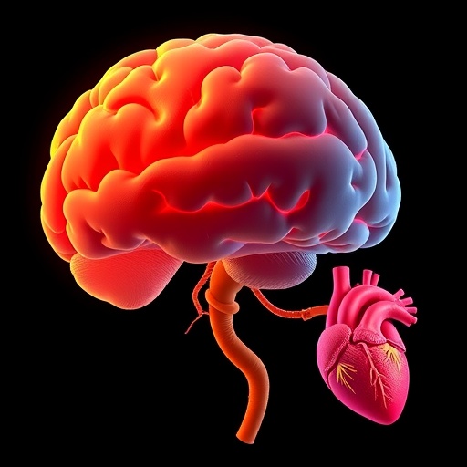
In a groundbreaking study published in the Journal of Perinatology this March, researchers have unveiled compelling evidence that fetuses diagnosed with congenital heart disease (CHD) exhibit significant variations in brain volume, intricately linked to abnormalities in placental vascular development. This insight marks a pivotal advancement in our understanding of fetal neurodevelopment and the complex interplay between the developing heart, brain, and placenta during gestation.
The brain undergoes rapid and critical growth phases throughout fetal development, a process that is delicately balanced by the oxygen and nutrient supply derived from both maternal and placental circulation. When congenital heart disease disrupts normal cardiovascular physiology, it alters this essential support system, potentially compromising cerebral maturation. Prior investigations have noted neurodevelopmental delays in children with CHD; however, direct correlations between the fetal brain volume and placental vascular architecture have remained elusive until now.
The study by O’Brien et al. employed advanced fetal magnetic resonance imaging (MRI) techniques to quantify brain volumes in a cohort of fetuses diagnosed prenatally with various forms of CHD. These volumetric scans were meticulously analyzed alongside detailed placental assessments, leveraging sophisticated imaging modalities capable of capturing subtle vascular anomalies. The multidisciplinary approach underlines the importance of integrating fetal cardiology, radiology, and perinatology to unravel complex pathophysiological mechanisms.
One of the most striking revelations from this research was the consistent observation of reduced total brain volumes in fetuses afflicted by CHD when compared to healthy controls. This reduction was not uniform but appeared to be influenced by the degree and type of placental vascular abnormalities detected. Notably, fetuses with more pronounced placental vascular disruption demonstrated more severe deficits in brain growth metrics, suggesting a dose-response relationship inherently linked to placental function.
The placenta serves as the crucial interface between mother and fetus, orchestrating gas exchange, nutrient delivery, and waste removal. Vascular abnormalities within this organ can result in compromised perfusion, exacerbating hypoxic conditions already burdened by impaired cardiac output in CHD cases. This dual-hit scenario potentially accelerates neurodevelopmental compromise. The findings challenge previous assumptions that CHD-related brain alterations arise solely from cardiac anomalies and highlight the placenta’s pivotal role in fetal brain growth.
From a mechanistic perspective, the study postulates that aberrant placental vascular remodeling interferes with the hemodynamic stability necessary for optimal cerebral oxygenation. In fetuses with CHD, inefficient blood flow and oxygen delivery may trigger compensatory but insufficient neuroprotective responses, culminating in structural brain changes observable via MRI. This pathophysiological model introduces new paradigms for understanding fetal brain injury and opens avenues for targeted therapeutic interventions.
Importantly, the research methodology demonstrated a rigorous design incorporating prospective enrollment, consistent imaging protocols, and control for confounding maternal factors such as preeclampsia or diabetes, which are known to independently affect placental health. The reproducibility and robustness of the imaging analysis reinforce the credibility of the findings and their potential applicability in clinical settings.
Clinicians engaged in prenatal care can leverage these insights to enhance risk stratification and counseling for expecting families confronted with a diagnosis of fetal CHD. By integrating placental assessment into routine fetal imaging protocols, it may become feasible to identify pregnancies at heightened risk for adverse neurodevelopmental outcomes earlier, enabling the consideration of interventions such as maternal oxygen therapy or optimized delivery planning.
Furthermore, this study propels a paradigm shift regarding the etiological narrative of neurodevelopmental delay in CHD, emphasizing that the brain’s vulnerability is a multifactorial phenomenon intricately tied to placental health rather than cardiac pathology alone. This holistic viewpoint may stimulate renewed research momentum focusing on modifiable placental factors within the intrauterine environment.
The broader implications of these findings extend to the design of future longitudinal studies evaluating neurocognitive trajectories postnatally. Early identification of placental vascular pathology could facilitate tailored neurodevelopmental surveillance and intervention programs, potentially mitigating long-term morbidity in this vulnerable population. Such proactive approaches represent the frontier of precision perinatal medicine.
In exploring translational prospects, the study hints at the potential for pharmacological or lifestyle interventions aimed at enhancing placental vascular function during critical windows of vulnerability. Complementary research into placental biomarkers may provide non-invasive routes to monitor fetal well-being and refine individualized care pathways.
Moreover, the technological advancements exemplified by the high-resolution fetal MRI and placental imaging protocols set new standards for fetal assessment. These imaging techniques could be expanded to study a spectrum of fetal conditions associated with compromised placental function, further enriching the field of fetal medicine.
While the results illuminate a novel dimension of fetal pathophysiology, they also underscore ongoing challenges. The complexity of fetal-placental-heart interactions necessitates sustained multidisciplinary collaboration and innovation. Additionally, ethical considerations regarding prenatal diagnosis and intervention remain critical discussions as such knowledge enters clinical practice.
In summary, the study by O’Brien and colleagues represents a transformative insight into the nexus of congenital heart disease, placental vascular health, and fetal brain development. This research not only advances scientific understanding but also lays the groundwork for enhancing prenatal diagnostic precision, therapeutic targeting, and ultimately improving neurodevelopmental outcomes for children born with CHD.
As science continues to decode the mysteries of fetal development, this landmark study accentuates the indispensable role of the placenta as partner and mediator in shaping brain growth amid cardiac challenges. The integration of advanced imaging, meticulous clinical evaluation, and physiological insight heralds a new era in fetal medicine—one in which the triad of heart, brain, and placenta is recognized as a unified system demanding comprehensive care and research focus.
Subject of Research:
Fetal brain development in the context of congenital heart disease and placental vascular abnormalities.
Article Title:
Brain volumes in fetuses with congenital heart disease and placental vascular abnormalities.
Article References:
O’Brien, E.A., Wypij, D., Rofeberg, V. et al. Brain volumes in fetuses with congenital heart disease and placental vascular abnormalities. J Perinatol (2026). https://doi.org/10.1038/s41372-026-02601-4
Image Credits:
AI Generated
DOI:
13 March 2026
Keywords:
Congenital heart disease, fetal brain volume, placental vascular abnormalities, fetal MRI, neurodevelopment, placental perfusion, prenatal diagnosis, fetal hypoxia, perinatal outcomes, placental pathology
