A groundbreaking study at Penn State University has unveiled new insights into the conductive polymer PEDOT:PSS, potentially revolutionizing implantable biomedical devices such as pacemakers and glucose monitors. Using cutting-edge cryogenic electron microscopy (cryo-EM), researchers led by Professor Enrique Gomez have decoded the complex nanostructure of this stretchy material widely employed in soft robotics and touchscreen technology. Their discovery, recently published in Nature Communications, highlights how subtle chemical modifications can dramatically enhance the polymer’s electrical and mechanical properties, opening promising avenues for bioelectronic innovation.
PEDOT:PSS is a unique conductive polymer that bridges the disparate electrical conduction mechanisms of human biology and electronic devices. Biological systems rely on ionic currents — essentially salt and ion mixtures — to transmit electrical signals, whereas electronic hardware uses electron flow through metal and semiconductors. The dual capability of PEDOT:PSS to conduct both electrons and interact sensitively with ionic currents makes it an extraordinary candidate for interfacing electronics with living tissue without compromising performance or biocompatibility.
Despite its ubiquity in emerging technologies, PEDOT:PSS has remained somewhat enigmatic at the molecular level until now. Gomez’s team employed cryo-electron microscopy to scrutinize the polymer’s structural assembly frozen at cryogenic temperatures, enabling visualization at near-atomic resolution. Unlike classical optical microscopes that rely on light and lenses, cryo-EM harnesses electron beams to reveal ultrastructural features, which has been transformative for virology and protein research and is now reshaping polymer science.
The experimental approach involved rapidly freezing meticulously prepared polymer samples embedded in nanoscopic films less than the width of a human hair. These samples included varied compositions with different salt additives to observe how chemical environment influences polymer morphology. By plunging them into liquid ethane at -180°C, the method preserved delicate molecular architectures against electron beam damage, allowing precise investigation of how salts promote the growth of conductive hairlike fibrils within the gel matrix.
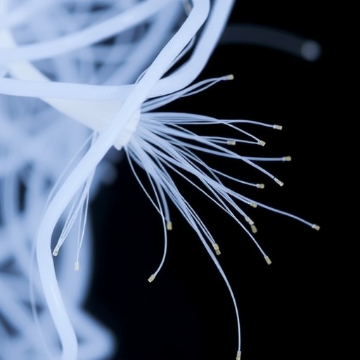
Researchers found that salt inclusion significantly elevated the formation of these whisker-like fibers, which act as highly efficient conduits for electrical charge transport. This direct correlation between salt additives and fiber density elucidates a previously uncharted mechanism for tuning conductivity in PEDOT:PSS. Crucially, the fibers persist even after the polymer absorbs water, maintaining electrical performance while imparting flexibility — a vital quality for implantable devices that must endure mechanical stress.
Water’s role emerged as equally critical. When hydrated, PEDOT:PSS transitions into a softer, more elastomeric gel that enhances stretchability without compromising conductivity. The study demonstrated that lithium salts increase the polymer’s affinity for water uptake, further promoting its mechanical resilience. Conversely, drying renders the material brittle, underscoring that water content is essential for the polymer’s durability and function in biointerfaces.
The dynamic interplay between ionic salts, water content, and polymer nanostructure reveals an intricate “templating” effect, whereby salts guide fiber assembly and dictate mechanical properties. This delicate balance results in a material that uniquely combines high stretchability with stable electrical conductivity — previously unattainable in synthetic polymers designed for bioelectronic applications. The implications extend beyond materials science into the realm of biomedical engineering, where durable, biocompatible conductors are desperately needed.
Professor Gomez envisions that these discoveries will inform next-generation designs for pacemakers that last longer, epidermal sensors with improved signal fidelity, and electromyography devices that better interface with muscles and nerves. The ability to finely control polymer microarchitecture through chemical additives marks a significant step toward personalized bioelectronic interfaces tailored for individual patient needs.
Looking ahead, the Penn State team plans to deepen their exploration of the chemical interactions driving fiber formation and conductivity enhancement. Identifying the fundamental molecular dynamics by which salts interact with polymer chains could unlock further improvements in material performance and inspire novel synthetic routes. Understanding these mechanisms is pivotal for scaling the technology and integrating it into commercial biomedical devices.
This research, supported by the U.S. National Science Foundation, the Office of Naval Research, and the National Institutes of Health, exemplifies the power of interdisciplinary collaboration, combining expertise in chemical engineering, materials science, chemistry, and life sciences. Co-authors from Penn State alongside partners from Iowa State University contributed to the comprehensive experimental design and analysis underpinning these findings.
Cryo-electron microscopy’s application in polymer science signals a transformative era in materials research, enabling unprecedented visualization of complex soft matter systems. Coupling molecular-level imaging with chemical manipulation offers a blueprint for designing next-generation functional materials with unparalleled combinations of electrical and mechanical properties suited for interfacing with living organisms.
By bridging the fundamental understanding of polymer nanostructure and practical material engineering, this study paves the way for high-performance, biocompatible materials crucial for advancing medical technology. As human-machine interfaces become more sophisticated and pervasive, innovations like those emerging from PEDOT:PSS research will be essential to the development of seamless, responsive bioelectronic systems that improve health outcomes and patient quality of life.
Subject of Research: Not applicable
Article Title: Cryogenic transmission electron microscopy reveals assembly and nanostructure of PEDOT:PSS
News Publication Date: 10-Feb-2026
Web References:
https://doi.org/10.1038/s41467-026-68890-7
References:
Gomez, E., et al. “Cryogenic transmission electron microscopy reveals assembly and nanostructure of PEDOT:PSS.” Nature Communications, 10 Feb. 2026. DOI: 10.1038/s41467-026-68890-7.
Image Credits: Ty Tkacik/Penn State
Keywords
Cryo electron microscopy, Biochemistry, Chemical engineering, Polymer chemistry, Polymer engineering, Engineering
