Despite decades of widespread immunization, whooping cough is resurging around the world, and a newly published review argues that the key to understanding why lies in a specific arm of the immune system: CD4+ T cells. The review, published in the journal Discover Immunity, synthesizes findings from animal models and human studies to explain how different pertussis vaccines and natural infection generate strikingly different T-cell responses, and why those differences determine whether the bacterium Bordetella pertussis is cleared from the respiratory tract or persists silently and spreads onward.
The scale of the resurgence is stark. In 2024, approximately 89 percent of infants globally received at least one dose of a diphtheria, tetanus, and pertussis-containing vaccine, and 85 percent completed at least three immunizations. Yet 941,565 confirmed pertussis cases were reported worldwide in 2024, a drastic increase from the 163,388 confirmed cases recorded in 2023. The rise became especially evident after the COVID-19 pandemic, with a substantial increase in global reported cases. Improved surveillance and more sensitive diagnostics have contributed to increased case detection, but they do not fully explain the trend.
The story begins with a vaccine switch. Whole-cell pertussis vaccines, which contain chemically inactivated B. pertussis bacteria, were introduced in the 1950s. Concerns over reactogenicity led many countries to replace them in the late 1990s with acellular vaccines composed of purified bacterial virulence factors. Since that switch, cyclic increases in pertussis cases have been observed worldwide. The acellular vaccine does protect against disease, likely through the induction of antibodies against pertussis toxin. But data from mice and nonhuman primate models show that acellular immunization fails to prevent nasal colonization, permitting subsequent transmission of the bacterium even among the vaccinated.
The immunological divide between the two vaccine types is central to the review. Natural infection and whole-cell vaccination both generate long-lasting TH1 and TH17 CD4+ T-cell responses. TH1 differentiation during infection is driven in part by interleukin-12 produced by airway macrophages responding to bacterial lipopolysaccharide, and interleukin-12 also stimulates interferon-gamma production from natural killer cells early in infection, reinforcing the TH1 polarization. In mice, CD4+ T cells are recruited to the lungs by day seven after challenge. Interferon-gamma from TH1 cells polarizes airway macrophages toward a state that kills B. pertussis through nitric oxide-dependent and independent mechanisms, and depleting these macrophages increases bacterial burden in the lungs. Interferon-gamma is required for efficient clearance of B. pertussis from the lung, and mice deficient in it show increased bacterial burden compared with wild-type animals.
TH17 responses, meanwhile, are critical at mucosal barrier sites. B. pertussis infection recruits T cells to the nasal cavity that produce interleukin-17, and these responses are essential for clearing infection from the nose. Bacterial components such as lipooligosaccharide, adenylate cyclase toxin, and pertussis toxin induce innate immune cells, including dendritic cells and macrophages, to produce the TH17-polarizing cytokines interleukin-1beta, interleukin-6, interleukin-21, and interleukin-23. Mice deficient in either interleukin-6 or interleukin-1beta show impaired bacterial clearance. Loss of interleukin-17 prevents clearance of B. pertussis from the nose entirely. TH17 cells promote chemokines that recruit monocytes and neutrophils, and Siglec-F-positive neutrophils in the nasal cavity deploy extracellular traps, reactive oxygen species, and phagocytosis against the bacterium. Depleting neutrophils impairs clearance in convalescent mice. The bacterium fights back: pertussis toxin and adenylate cyclase toxin accumulate intracellular cyclic AMP in immune cells, inhibiting oxidative burst, extracellular trap formation, and phagocytosis.
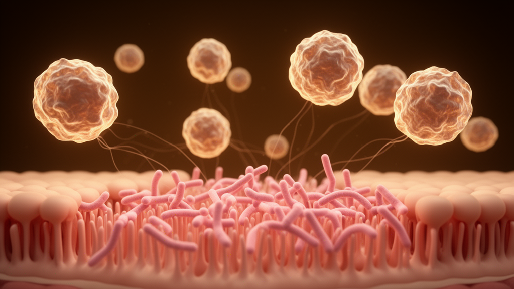
Perhaps the most consequential players are tissue-resident memory T cells, or TRM cells. These long-lived cells, generated by natural infection, reside in tissues and do not circulate through the blood, providing a rapid first line of defense at the site of re-exposure. In mouse models, CD4+ TRM cells appear in the lungs as early as one to two weeks after infection and persist in the lungs and nose after the bacterial load is eliminated. They are detected as long as seven months after bacterial challenge is cleared. In the lungs they produce primarily interferon-gamma; in the nose they produce primarily interleukin-17. Upon reinfection, TRM cells rapidly expand locally and are essential for reducing bacterial burden in both sites. In a striking experiment, convalescent mice treated with the drug FTY720, which traps circulating lymphocytes in lymph nodes and prevents them from reaching the infection site, still showed expansion of mucosal TRM cells and reduced bacterial load in the lungs, suggesting that tissue-resident cells alone can eliminate the bacteria. Adoptive transfer of TRM cells from convalescent mice into naive mice likewise decreased bacterial burden in the recipients.
Whole-cell vaccination mirrors these protective features. Intramuscular immunization with whole-cell vaccine induces TH1/TH17-polarized systemic responses that are drawn to the respiratory mucosa upon challenge, establishing interferon-gamma-producing TRM cells in the lungs and interleukin-17-positive TRM cells in the nose. In wPV-immunized mice, bacterial load in the nose is reduced to the limit of detection, and studies in baboons showed that wPV-immunized animals do not transmit the infection. Acellular vaccination produces a very different picture: robust systemic TH2 responses driven by interleukin-4, producing interleukin-4, interleukin-5, and interleukin-13. While both vaccine types generate strong IgG antibody responses, children vaccinated with an acellular alum-adsorbed pertussis toxoid show mainly IgG1 and IgG4 against pertussis toxin, whereas natural infection and whole-cell immunization elicit IgG1 and IgG3. Nonhuman primate and human studies show that antibody titers and protective immunity wane more rapidly after acellular vaccination, and epidemiological data indicate that aPV-mediated protection can decline substantially within four years of a booster dose. One proposed explanation involves follicular T helper cells: mice immunized with whole-cell vaccine generated more of these follicular helper cells and higher levels of the chemokine CXCL13, which persisted longer in lymph nodes and supported B-cell recruitment, whereas follicular helper populations in acellular-vaccinated mice dropped drastically 60 days after immunization even though antibody levels remained elevated for 532 days.
Acellular vaccination also induces regulatory T cells that may blunt protective immunity. Antigen-specific CD4+ regulatory T cells producing interleukin-10, interferon-gamma, and interleukin-5 suppress interleukin-17 production by TH17 cells, and acellular immunization additionally induces interleukin-10-producing CD8+ regulatory T cells that suppress both TH1 and TH17 activation. Blocking interleukin-10 after acellular immunization increased CD4+ TRM cells expressing interleukin-17. The review’s authors suggest the immune response generated by the acellular vaccine is suboptimal and less durable, which may underlie its inability to prevent nasal colonization. Compounding the problem, circulating clinical strains increasingly lack key vaccine antigens, including filamentous hemagglutinin, pertactin, and pertussis toxin, and pertactin-deficient strains continue to increase in countries using acellular vaccines, though they have also been isolated where whole-cell vaccination remains standard. These antigens are components of the current acellular vaccine, suggesting vaccine-driven selection may contribute.
Emerging strategies aim to close the gap. Researchers are screening for conserved antigens less susceptible to antigenic modulation, including through Tnseq screens in nonhuman primates and whole-genome mapping of CD4+ T-cell responses in immunized humans. Novel adjuvants that polarize responses toward TH1 and TH17 are being paired with mucosal delivery routes: a TLR2 agonist called LP1569 decreased bacterial burden in the lower respiratory tract; adding the TLR4 agonist BcfA to an approved acellular vaccine, delivered by intramuscular priming followed by an intranasal booster, decreased bacterial burden in the lungs and nasal cavity alongside increased TH1/TH17 TRM cells; and TLR9 agonist CpG ODN, STING agonist c-d-GMP, curdlan, and the OMV-based adjuvant T-vant have each shown promise in mouse studies. A live-attenuated intranasal vaccine, BPZE1, elicits antibody and TH1/TH17 responses resembling those of the reference whole-cell vaccine, protected mice one year after immunization, reduced nasal colonization in baboons, and has been shown to be safe, with ongoing clinical trials evaluating immune responses and protective efficacy. The authors caution that most mechanistic work has been done in mice, that human mucosal tissues such as tonsils will be instrumental in validating these findings, and that further studies are needed to determine whether the magnitude, specificity, and longevity of CD4+ T-cell responses can serve as correlates of protection. Maternal acellular vaccination protects neonates before their own immunization, and improved maternal vaccines could extend that protection. Safety evaluation after mucosal delivery, manufacturing feasibility, and regulatory hurdles for mucosal subunit vaccines remain significant considerations before any next-generation vaccine reaches the clinic.
Subject of Research: CD4+ T-cell immunity to Bordetella pertussis and implications for next-generation pertussis vaccines
Article Title: CD4+ T-cell immunity to Bordetella pertussis
Article References: Hall, J. M., Defalco, J. B., & Dubey, P. (2026). CD4+ T-cell immunity to Bordetella pertussis. Discover Immunity, 3(1), Article 12. https://doi.org/10.1007/s44368-026-00030-1
Image Credits: AI Generated
DOI: 10.1007/s44368-026-00030-1
Keywords: Bordetella pertussis, whooping cough, CD4+ T cells, TH1/TH17, tissue-resident memory T cells, acellular pertussis vaccine, whole-cell pertussis vaccine, mucosal immunity, vaccine resurgence, T regulatory cells, intranasal vaccination, pertussis toxin
Cite Scienmag News
Kristina Jarvis. (October 29, 2026). Why Whooping Cough Is Coming Back: T Cells Hold the Answer. Scienmag. https://scienmag.com/why-whooping-cough-is-coming-back-t-cells-hold-the-answer/
Kristina Jarvis. "Why Whooping Cough Is Coming Back: T Cells Hold the Answer." Scienmag, 29 October 2026, https://scienmag.com/why-whooping-cough-is-coming-back-t-cells-hold-the-answer/. Accessed 29 October 2026.
Kristina Jarvis. "Why Whooping Cough Is Coming Back: T Cells Hold the Answer." Scienmag. October 29, 2026. https://scienmag.com/why-whooping-cough-is-coming-back-t-cells-hold-the-answer/