The advent of three-dimensional histology has opened unprecedented avenues for exploring the complex molecular landscapes within intact organs. Traditionally, biological inquiry at the cellular and tissue levels has relied heavily on two-dimensional histological sections, which inherently limit spatial context and molecular insight. Breaking through these constraints, the innovative technique known as Tris buffer-mediated retention of in situ hybridization chain reaction signal in cleared organs, abbreviated as TRISCO, now promises to transform the field of spatial transcriptomics by enabling comprehensive three-dimensional RNA mapping at single-cell resolution across entire organs.
One of the longstanding challenges in molecular histology has been the reliable visualization of RNA transcripts throughout large volumes of tissue without compromising spatial integrity or molecular fidelity. Existing methods have predominantly focused on protein imaging, leveraging antibodies and fluorescent labeling to reveal cellular architecture and protein distribution. However, RNA—the direct blueprint of gene expression—has remained elusive in whole-organ analysis due to issues such as signal diffusion, incomplete penetration, or signal loss during chemical treatments. TRISCO addresses these challenges head-on with a buffer-mediated retention strategy that preserves RNA hybridization signals effectively during tissue clearing, ensuring homogenous and stable labeling throughout volumetric samples.
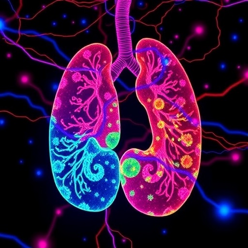
The TRISCO protocol is ingeniously straightforward yet meticulously optimized to maintain signal integrity through multiple steps that integrate hybridization chain reaction (HCR) fluorescent amplification within cleared organs. This method employs a Tris-buffered environment that stabilizes the in situ HCR probes and fluorophores during the clearing process, circumventing common pitfalls like signal quenching or uneven penetration. By doing so, TRISCO dramatically enhances the reliability of RNA visualization in intact murine organs, including the brain, lung, heart, kidney, and spinal cord. Its versatility further extends to larger rodent models such as rat and guinea pig brains, underscoring its broad applicability.
A defining feature of TRISCO lies in its adaptability and accessibility. The protocol avoids reliance on specialized instruments or harsh chemical treatments that may perturb tissue architecture or require extensive expertise. Instead, it leverages standard laboratory reagents and equipment, making it approachable for a wide spectrum of researchers engaged in neuroscience, developmental biology, or translational medicine. The entire workflow, from sample preparation through hybridization and imaging, can be completed within 10 to 15 days, with flexible pause points that accommodate routine scheduling constraints common in experimental research environments.
At the core of TRISCO’s success is its ability to maintain the spatial fidelity of RNA transcripts throughout the clearing process. Traditional tissue clearing methods, while excellent at rendering tissues optically transparent, often engender the loss or redistribution of molecular labels due to chemical exposure or prolonged enzymatic digestion. TRISCO’s Tris-buffer system creates a protective milieu that locks hybridization chain reaction signals in place, preserving delicate RNA structures within the native tissue context. This advancement allows for confident assignment of RNA transcripts to their correct cellular environments in three dimensions.
Light-sheet fluorescence microscopy synergizes impeccably with the TRISCO method, offering rapid volumetric imaging with reduced photobleaching and phototoxic effects compared to confocal microscopy. The clarity and homogeneity of fluorescent signals retained via TRISCO empower researchers to capture exquisite 3D reconstructions of gene expression patterns across whole organs. These reconstructions can resolve individual cells’ transcriptomic states while simultaneously mapping their precise anatomical locations, a feat unattainable with conventional 2D histology or lower-resolution imaging approaches.
The implications of TRISCO’s capability ripple through numerous fields of biological research. Neuroscientists, for instance, gain the ability to decode complex cellular networks and transcriptional states underpinning brain function and pathology in unprecedented detail. Likewise, developmental biologists can map dynamic changes in gene expression throughout organogenesis or tissue regeneration, observing the interplay between different cell populations within intact environments. In translational contexts, the technology facilitates the study of disease models in ways that preserve the spatial nuances critical to understanding pathophysiology.
TRISCO’s utilization across diverse organs confirms its robustness and reproducibility. Its application in mouse lung tissues elucidates respiratory cell heterogeneity and gene regulation under both normal and diseased conditions. In cardiac tissue, TRISCO enables the visualization of transcriptional profiles in cardiac myocytes, fibroblasts, and endothelial cells within the three-dimensional organ matrix, informing studies of heart disease and repair. The kidney and spinal cord further demonstrate TRISCO’s versatility by revealing regional transcriptomic signatures vital for organ-specific function and responses.
Beyond biological insight, TRISCO’s user-friendly design lowers entry barriers for laboratories lacking high-end expertise or equipment for complex spatial transcriptomic approaches. The protocol’s straightforwardness ensures that emerging labs or interdisciplinary teams can adopt and modify it according to their experimental needs. The elimination of harsh chemical treatments also safeguards tissue integrity, facilitating downstream analyses or multimodal imaging, including combining RNA visualization with protein or structural markers.
As spatial transcriptomics continues to ascend as a transformative technology, methods like TRISCO are essential for its evolution toward whole-organ, high-resolution applications. The capacity to visualize single-molecule RNA distributions throughout cubic millimeters of intact tissues can reveal subtle cellular heterogeneities, regulatory networks, and microenvironmental influences previously masked in dissected or dissociated samples. This holistic molecular cartography carries profound ramifications for both basic and clinical sciences.
Looking ahead, integration of TRISCO with emerging multiplexed RNA detection chemistries, machine learning-driven image analysis, and multi-omics pipelines promises to exponentially expand the depth and breadth of spatial molecular profiling. Investigators may soon achieve simultaneous, ultra-high-resolution mapping of transcripts, proteins, and metabolites across whole organs, shedding new light on the interplay between genotype, phenotype, and environment in health and disease. The groundwork laid by TRISCO is thus a cornerstone of the future molecular anatomy toolkit.
In summary, TRISCO introduces a powerful, accessible, and versatile platform for in situ hybridization chain reaction-based RNA visualization in cleared tissues, enabling true single-cell-resolution spatial transcriptomics at the whole-organ scale. By preserving thorough, homogeneous labeling and compatibility with light-sheet microscopy, this method emerges as a vital facilitator of next-generation 3D histological and molecular investigations. Its potential to revolutionize our understanding of complex biological systems makes TRISCO a landmark advancement with far-reaching impact across biomedical research disciplines.
As spatially resolved transcriptomics accelerates toward clinical translation, TRISCO’s ability to provide comprehensive molecular maps without compromising tissue integrity or accessibility positions it uniquely for use in disease modeling and therapeutic discovery. The balance of technical simplicity with sophisticated molecular preservation captures an ideal intersection that will empower researchers worldwide to unravel the intricacies of cellular organization in intact biological systems. The gene expression atlases enabled by TRISCO promise to become foundational resources charting the spatial logic of biological function and dysfunction.
The development and dissemination of TRISCO leader researchers highlights the importance of cross-disciplinary innovation bridging molecular biology, chemistry, microscopy, and computational analysis. Its broad adoption will redefine how spatial transcriptomic data is generated and interpreted, catalyzing scientific breakthroughs from basic cell biology to personalized medicine. This technology heralds a new era where observing RNA landscapes in whole organs is accessible, reliable, and scalable, inspiring a wave of discoveries that will reshape our biological understanding and capabilities.
Subject of Research: Whole-organ spatial transcriptomic analysis at single-cell resolution.
Article Title: Whole-organ spatial transcriptional analysis at cellular resolution using TRISCO.
Article References:
Li, Y., Walton, A., Kreutzmann, J.C. et al. Whole-organ spatial transcriptional analysis at cellular resolution using TRISCO. Nat Protoc (2026). https://doi.org/10.1038/s41596-026-01386-2
DOI: https://doi.org/10.1038/s41596-026-01386-2
