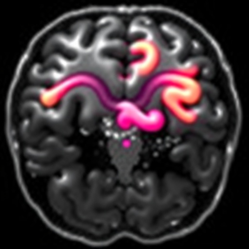
A New Diagnostic Frontier: Punctate White Matter Lesions as a Harbinger of Cerebral Palsy in Preterm Infants
In the rapidly evolving field of neonatal neurology, early and accurate prediction of neurodevelopmental outcomes remains a paramount challenge. Recent groundbreaking research has shed light on a subtle but significant neuroimaging biomarker—punctate white matter lesions (PWML)—which may hold the key to predicting the risk of cerebral palsy in preterm infants with remarkable precision. This work, emerging from a collaboration between neonatal neurologists and radiologists, underscores the urgent need for revising current neonatal imaging protocols to incorporate routine brain magnetic resonance imaging (MRI) in this vulnerable population.
PWML, characterized by small, focal abnormalities detectable on MRI within the cerebral white matter, have historically been regarded with some ambiguity in their clinical significance. These lesions appear as discrete hyperintensities on T1-weighted images and hypointensities on T2-weighted sequences, scattered throughout delicate white matter tracts. Their pathophysiology is believed to stem from microvascular disruption and localized glial injury due to the relative fragility of the immature cerebral vasculature in preterm neonates between 24 and 32 weeks of gestational age. Until now, the association between PWML and long-term neurodevelopmental deficits, including motor impairment, had been suggested but remained inconclusive.
The compelling findings from the latest study definitively correlate the presence and distribution of PWML with an increased risk of cerebral palsy, a lifelong movement disorder resulting from early brain injury. Pediatric neurologists have long sought reliable prognostic markers that could facilitate early intervention and supportive therapy to mitigate the devastating sequelae of cerebral palsy. By utilizing advanced neuroimaging protocols combined with rigorous longitudinal clinical follow-up, clinicians now have tangible evidence that routine brain MRI in preterm infants can provide critical prognostic information beyond conventional cranial ultrasound.
The implications of integrating routine MRI scanning into neonatal intensive care units (NICUs) are profound. Unlike ultrasound, which often fails to detect the subtle white matter abnormalities characteristic of PWML, MRI offers unparalleled spatial resolution and tissue contrast, enabling the visualization of these minute lesions. Early identification of infants harboring PWML could lead to targeted neuroprotective strategies, including optimized respiratory support, neurorehabilitation, and potentially pharmacologic interventions aimed at minimizing secondary neuronal injury.
Importantly, this research leverages a multidisciplinary approach utilizing neuroradiology, neonatal neurology, and developmental pediatrics to build a robust correlation between lesion burden, lesion location, and subsequent motor outcome severity. Lesions localized to periventricular and subcortical regions, areas subserved by critical motor pathways, were especially predictive of later diagnosis of spastic diplegia, one of the most common clinical manifestations of cerebral palsy in prematurity. The stratification of lesion topography offers a nuanced predictive model previously unavailable in neonatal neuroimaging practice.
From a technical standpoint, the study deployed advanced MRI sequences, including diffusion-weighted imaging (DWI) and susceptibility-weighted imaging (SWI), to enhance lesion detectability. DWI is sensitive to cytotoxic edema, often preceding irreversible tissue damage, while SWI highlights microhemorrhages that often accompany PWML. These modalities, combined with refined segmentation algorithms, allowed for quantification of lesion volume and mapping onto white matter tracts, correlating structural abnormalities with neurofunctional outcomes.
Beyond clinical practice, this new understanding of PWML contributes to the fundamental neuroscience of prematurity-related brain injury. It supports the hypothesis that cerebral white matter damage, a hallmark of encephalopathy of prematurity, encompasses a spectrum wherein punctate lesions represent focal ischemic insults. These, in turn, disrupt oligodendrocyte maturation and myelination, critical processes during late gestation brain development. The interplay between vascular insult and glial vulnerability elucidated by this work deepens our grasp of the pathobiology underpinning cerebral palsy.
While the study unequivocally supports the inclusion of routine MRI in the clinical evaluation of preterm infants, logistical and economic challenges must be addressed. MRI requires specialized equipment and sedation protocols that pose risks and limitations in the fragile neonatal population. Nevertheless, advances in quiet, motion-robust imaging technologies and rapid sequence acquisitions are making bedside-compatible neonatal MRI a realistic goal. Health systems must consider these investments justified given the potential long-term cost savings by enabling earlier, individualized therapeutic interventions.
From a public health perspective, this research has the potential to transform neonatal care paradigms globally. Standardized neuroimaging protocols including routine MRI could become part of evidence-based guidelines, fostering equitable access to high-quality diagnostic evaluation for at-risk infants. Early detection would also empower families and caregivers with prognostic clarity and facilitate enrollment in early intervention programs that improve neurodevelopmental trajectories.
The impact of this research may ripple into future therapeutic trials, informing inclusion criteria and serving as an objective biomarker for treatment response. For example, neuroprotective agents targeting inflammation and oxidative stress pathways could be stratified based on lesion presence and severity, refining precision medicine approaches in neonatal neurology. Moreover, PWML quantification and localization may serve as surrogate endpoints, accelerating the pace of clinical innovation.
Importantly, this landmark study highlights the indispensable role of longitudinal follow-up in correlating imaging findings with clinical outcomes. Multidisciplinary teams performed serial developmental assessments extending into early childhood, ensuring that MRI markers are not only cross-sectional snapshots but predictive tools of functional prognosis. This comprehensive approach sets a new standard in neonatal neurocritical care research.
Ethical considerations also emerge from the implementation of routine MRI screening, including informed consent, the psychological impact of early risk notification on families, and managing incidental findings unrelated to cerebral palsy risk. Neonatal care providers will need training to navigate these complex conversations compassionately while maintaining transparency and evidence-based counseling.
Looking ahead, integration of artificial intelligence and machine learning algorithms promises to enhance the sensitivity and specificity of PWML detection. Automated lesion segmentation and risk modeling could reduce diagnostic variability and augment clinical decision-making. The convergence of big data analytics with neonatal neuroimaging heralds an exciting era of personalized neurodevelopmental care.
In conclusion, the identification of punctate white matter lesions as potent predictors of cerebral palsy risk marks a significant advancement in neonatal neurology. This breakthrough not only challenges existing screening paradigms but also opens new horizons for early intervention and improved outcomes for the most vulnerable infants. As research continues to dissect the complexities of prematurity-related brain injury, routine brain MRI stands out as an indispensable tool — a lens into the developing brain that promises hope amidst uncertainty.
Subject of Research: Prediction of cerebral palsy risk in preterm infants through detection of punctate white matter lesions via routine brain MRI.
Article Title: Punctate white matter lesions predict risk for cerebral palsy: further evidence for routine brain MRI in preterm infants.
Article References:
Selvanathan, T., Gano, D. Punctate white matter lesions predict risk for cerebral palsy: further evidence for routine brain MRI in preterm infants. Pediatr Res (2025). https://doi.org/10.1038/s41390-025-04333-1
Image Credits: AI Generated


