A simple blow into a handheld peak flow meter has long been promoted as a quick window into lung health, and a new analysis of two of the world’s largest aging studies now confirms that a weak reading really does foreshadow a doctor-diagnosed lung condition. But the same research delivers a sobering caveat: when peak expiratory flow is measured against the gold-standard numbers produced by full spirometry, it adds almost nothing that those richer measurements do not already capture. The findings, published in PLOS One, offer both reassurance about the signal and a caution about overselling the humble peak flow meter.
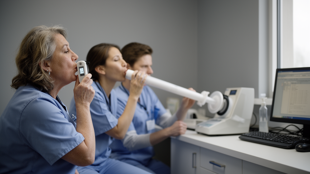
The study, led by Haiyang Hu and colleagues, drew on the Health and Retirement Study (HRS) in the United States and the English Longitudinal Study of Ageing (ELSA), two nationally representative cohorts that follow thousands of older adults for years, tracking their health, function, and self-reported diagnoses. Peak expiratory flow, or PEF, records the maximum speed at which a person can force air out of their lungs, expressed in liters per minute. It is cheap, fast, and requires only a small plastic device, which is why it appears in large population surveys where full spirometry equipment and trained technicians would be impractical. The question the researchers posed was deceptively simple: if an older adult records an unusually low PEF at one study visit, are they more likely to report a recognized lung disease, such as chronic obstructive pulmonary disease or asthma, for the first time at a later visit?
Answering that question rigorously required careful design choices that distinguish this analysis from many earlier studies of lung function screening. The team defined their outcome precisely as the first subsequently observed self-report of a recognized lung disease, not the biological onset of disease itself, an important distinction because people can carry airway pathology for years before it prompts a diagnosis or even a mention to a survey interviewer. They also adopted a person-disjoint replication strategy, meaning the statistical model was developed in one set of HRS participants and then tested, unchanged in its essential specification, in a completely separate set of people from the same study, and then again in the entirely independent ELSA cohort from another country. This guards against the common trap of overfitting, where a model looks impressive on the data that shaped it but collapses on new data.
The numbers behind the analysis are substantial. In the HRS discovery sample, the models spanned 18,510 person-windows of observation, during which 320 participants first reported a recognized lung disease. The prespecified replication sample within HRS contributed 11,424 windows and 188 first reports, while ELSA added 8,634 person-waves with 95 first reports. In each cohort, low PEF was defined slightly differently: in HRS, the researchers prespecified a nominal lower-decile z score, while in ELSA they used an empirically derived lower decile. Participants with low PEF were compared with peers using models that adjusted for demographic characteristics, clinical conditions, and functional status, with clustering at the participant level to account for repeated observations.
The central result was strikingly consistent across all three samples. In the HRS discovery data, low PEF was associated with roughly a threefold increase in the odds of a subsequent first lung-disease report, with an odds ratio of 3.36 and a 95 percent confidence interval of 2.54 to 4.45. The person-disjoint HRS replication yielded an odds ratio of 3.14 (2.19 to 4.49), and ELSA, an entirely different cohort in a different country, produced 3.77 (2.36 to 6.01). Sensitivity analyses using survey weighting produced similar estimates, strengthening the case that the association is not an artifact of sampling design. For a cheap, thirty-second test, that is a robust and portable signal.
Yet the study’s most revealing findings came from its comparisons and negative controls. The researchers included a stroke comparator as a sensitivity analysis, reasoning that if low PEF were merely a general marker of frailty or declining health rather than something specific to the respiratory system, it should predict stroke reports just as strongly. Instead, the stroke associations were weaker or null, suggesting that the PEF signal is relatively specific to lung disease rather than a generic alarm for bodily decline. This kind of disease-specificity check is often missing from biomarker studies, and its inclusion here adds credibility to the respiratory interpretation.
The picture grew more complicated when PEF was benchmarked against spirometry. In a post-hoc analysis within ELSA, the team compared PEF with two spirometric indices: forced expiratory volume in one second (FEV1), which measures how much air can be expelled in the first second of a forced breath, and forced vital capacity (FVC), the total volume of air exhaled. These measurements require the participant to blow into a calibrated spirometer through multiple maneuvers and are the standard diagnostic tools for obstructive and restrictive lung disease. After adjusting for separate FEV1 and FVC indices, the association between continuous PEF and first lung-disease report shrank to an odds ratio of 1.04 with a confidence interval of 0.90 to 1.20, comfortably spanning the null. In other words, once spirometry was accounted for, PEF retained no clearly detectable independent association.
Predictive performance told the same story. Among 1,584 participants in a fixed-horizon prediction exercise, of whom 95 experienced a first report, a model based on covariates alone achieved an area under the curve (AUC) of 0.650. Adding PEF nudged that to 0.684, a modest gain. Adding FEV1 and FVC lifted the AUC to 0.745, and combining all three measurements produced 0.739, no better than spirometry alone. Full-pipeline bootstrap sensitivity analyses, which repeated the entire modeling procedure many times to quantify uncertainty, found no clear incremental evidence for PEF beyond FEV1 and FVC. In exploratory fixed out-of-fold predictions, PEF did improve average precision and prediction error relative to covariates alone, but that advantage evaporated once the spirometric indices entered the model.
There is also a practical screening problem with any low-PEF rule. Across differing prediction horizons, thresholds flagging the lowest decile of peak flow identified fewer than one-third of the people who went on to report a recognized lung disease in both the HRS replication sample and ELSA. A screening test that misses more than two-thirds of future cases, even at a deliberately low threshold, has limited value as a stand-alone population tool, however strong its odds ratios may be at the group level. The authors are careful to note another conceptual point: because the outcome was the first observed self-report rather than biological onset, the association may partly reflect the process by which symptoms come to medical attention and get recorded, not a purely physiological pathway. Same-protocol collection of PEF and diagnoses in a survey, they emphasize, does not by itself establish a peak-flow-only biological route to disease.
What, then, should clinicians, survey designers, and the public take away? For large-scale aging surveys and resource-limited settings where spirometry is unavailable, the study validates peak flow as a meaningful, replicable marker: older adults with readings in the lowest decile face roughly three times the odds of a subsequent first lung-disease report, consistently across two countries and independent samples. That signal deserves attention and could justify closer respiratory follow-up. But where spirometry is feasible, the evidence points firmly toward FEV1 and FVC as the measurements that matter, both for association and for prediction. The peak flow meter earns its place as a useful sentinel, not as a substitute. As the population ages and chronic lung disease imposes a growing burden worldwide, the study is a reminder that the cheapest test and the best test are not always the same, and that rigorous replication and benchmarking, unglamorous as they are, remain the surest way to tell them apart.
Subject of Research: Association of low peak expiratory flow with subsequent first self-reported lung disease in older adults, benchmarked against spirometry
Article Title: Low peak expiratory flow and subsequent first self-report of recognized lung disease: Person-disjoint replication and spirometric benchmarking in HRS and ELSA
Article References: Hu, H., Wang, C., Fu, J., & Zhou, H. (2026). Low peak expiratory flow and subsequent first self-report of recognized lung disease: Person-disjoint replication and spirometric benchmarking in HRS and ELSA. PLOS One, 21(10), e0360668. https://doi.org/10.1371/journal.pone.0360668
Image Credits: AI Generated
DOI: 10.1371/journal.pone.0360668
Keywords: peak expiratory flow, spirometry, lung disease, Health and Retirement Study, English Longitudinal Study of Ageing, FEV1, FVC, COPD, aging cohorts, prediction, replication, epidemiology
Cite Scienmag News
Barbara Leach. (October 15, 2026). Weak Breath Test Flags Lung Disease Risk, But Spirometry Does It Better. Scienmag. https://scienmag.com/weak-breath-test-flags-lung-disease-risk-but-spirometry-does-it-better/
Barbara Leach. "Weak Breath Test Flags Lung Disease Risk, But Spirometry Does It Better." Scienmag, 15 October 2026, https://scienmag.com/weak-breath-test-flags-lung-disease-risk-but-spirometry-does-it-better/. Accessed 15 October 2026.
Barbara Leach. "Weak Breath Test Flags Lung Disease Risk, But Spirometry Does It Better." Scienmag. October 15, 2026. https://scienmag.com/weak-breath-test-flags-lung-disease-risk-but-spirometry-does-it-better/