


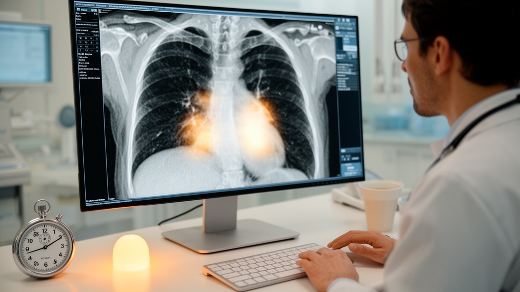
Epicardial adipose tissue, the fat layer that sits directly on the surface of the heart between the myocardium and the visceral layer of the pericardium, has moved steadily from anatomical curiosity to recognized cardiovascular signal. It is an active endocrine organ that exerts inflammatory effects on the coronary arteries and the heart as a whole, and population-based studies have linked larger volumes of it to obesity, diabetes mellitus, hypercholesterolemia and hypertension. It is also implicated in the development of coronary artery disease through the promotion of local inflammation. Measuring it, however, has traditionally demanded either echocardiography or, with superior accuracy, ECG-triggered cardiac computed tomography combined with specialized segmentation software, which keeps the measurement out of reach for many routine clinical settings.
A new study published in European Radiology Cardiovascular and Thoracic proposes a strikingly low-tech answer. Researchers at a single center developed a qualitative visual score that estimates epicardial adipose tissue volume directly from non-contrast, ECG-triggered cardiac CT scans, the kind of calcium-scoring acquisition many heart transplant recipients already undergo each year. In their retrospective cohort of 155 adult heart transplant recipients, the visual score correlated with automated volumetric measurements with a Spearman rank correlation coefficient of 0.965, a very strong association, and it could be applied in roughly one minute per scan after limited training.
The clinical motivation is particular to transplant medicine. Chronic immunosuppressive therapy after heart transplantation contributes considerably to obesity and type 2 diabetes mellitus, both risk factors for larger epicardial fat volumes. Evidence also indicates that patients experiencing a higher number of rejection episodes tend to exhibit greater epicardial adipose tissue accumulation, with fat volume progressively increasing with each rejection episode. The suspected mechanism involves the role of this fat in the development and progression of cardiac allograft vasculopathy, the leading cause of graft failure and mortality following transplantation. Monitoring the fat layer may therefore offer a window into cardiovascular risk, graft health and long-term survival in this population.
The study team, led by B. C. J. van Dijk with co-authors including D. Bos and O. C. Manintveld, included heart transplant recipients who were more than four years post-transplant and underwent CT imaging for cardiac allograft vasculopathy surveillance between February 2018 and June 2020. All scans were acquired on second- or third-generation dual-source CT scanners, with a non-enhanced cardiac CT protocol at 120 kVp and 3-mm slice thickness. Patients with impaired renal function, iodine allergy or simultaneous organ transplantation were excluded, as were scans in which scoring was not attainable. The median age at transplantation was 44 years, 34 percent of patients were female, and the median interval between transplantation and CT acquisition was 11 years.
The reference measurement came from a validated, custom-made automated algorithm developed by Shahzad and colleagues, which delineates the pericardium and counts all voxels with attenuation values between minus 200 and minus 30 Hounsfield Units within it as epicardial adipose tissue. That automated approach has shown excellent agreement and reproducibility against manual annotations by experienced radiologists, but it is not quick in practice. The workflow requires scan pre-processing, running the segmentation algorithm, and manual inspection and correction when necessary, so the time from scan acquisition to a reliable volumetric result was typically four to eight hours per scan.
The visual alternative works very differently. Observers score axial CT images at three predefined anatomical levels: the first slice showing the right atrial appendage, the level of the aortic valve, and the level where the inferior vena cava separates from the right atrium. At each level, the amount of fat is graded on a four-point scale from minimal to maximal, with scoring reflecting the overall fat burden on each slice whether the tissue is distributed focally in a limited area or dispersed diffusely around the heart. The three sub-scores are added to yield a total between 3 and 12. Observers trained for approximately one day using reference images drawn from a separate transplant cohort at the same center, deliberately kept out of the analysis dataset to avoid overlap between training material and study scans.
The results showed a stepwise increase in absolute fat volume across the visual score categories. Median measured volume ranged from 90.0 milliliters in patients with a total visual score of 3 up to 451.2 milliliters in those with a score of 12, with the number of patients per scoring category ranging from 4 to 31. A discrepancy between the qualitative score and the absolute volume appeared in only 4 of 155 patients, about 3 percent, in one case higher than expected and in three cases lower. Intra-observer agreement was excellent, with a weighted Cohen’s kappa of 0.81, and inter-observer agreement ranged from substantial to excellent, with kappa values of 0.66 and 0.81 for the two independent observers.
Notably, the method held up under imperfect imaging conditions. Thirty-one of the 155 scans, about 20 percent, contained relevant artifacts, including pacemaker leads in 27 cases, coronary stents in 3, and one stack artifact, and 2 scans showed unusual anatomical features. Every scan with artifacts or anatomical variation remained assessable with the visual score, an important property in a post-transplant population characterized by altered cardiac anatomy, scar tissue and hardware. The authors also caution that the method is not suitable for contrast-enhanced CT, because opacification of adjacent cardiac structures hampers accurate tissue discrimination by the human eye.
The study sits within a broader effort to make fat quantification practical. Earlier approaches include a threshold-based three-dimensional segmentation method applied to contrast-enhanced coronary CT angiography that took about five minutes per scan, and a deep-learning framework trained and validated on contrast-enhanced, ECG-triggered datasets whose performance in post-transplant anatomy remains uncertain. Quantitative methods on non-contrast scans have required substantial user interaction, with manual pericardial contour delineation taking roughly 7 to 11 minutes per scan, and clinical implementation of such quantitative techniques has remained limited by usability and cost. The visual score requires no contrast, no proprietary software and no additional imaging, since it runs on scans already acquired for calcium scoring.
The authors are careful about the boundaries of their findings. The study was retrospective and single-center, the population consisted exclusively of heart transplant recipients, and no external validation was performed, so reproducibility across institutions and applicability to broader patient groups remain open questions. Performance on non-ECG-triggered routine chest CT scans has not yet been determined, although the authors suggest lung and liver transplant follow-up imaging and lung cancer screening as realistic future settings to test. They also note that photon-counting CT with virtual non-contrast reconstructions may eventually enable reliable fat evaluation even within contrast protocols. For now, the reported message is narrower but concrete: in this cohort, a trained observer could estimate epicardial adipose tissue on a routine non-contrast cardiac CT in about a minute, with a correlation close to automated volumetry and agreement ranging from substantial to excellent, offering a practical surrogate where dedicated software or contrast-enhanced imaging is not available.
Subject of Research: A visual CT scoring method for estimating epicardial adipose tissue volume in heart transplant recipients
Article Title: A simple visual score to estimate epicardial adipose tissue volume on non-contrast ECG-triggered cardiac CT post-heart transplantation
Article References: van Dijk, B. C. J., Roest, S., de Boer, R. A., Budde, R. P. J., Manintveld, O. C., & Bos, D. (2026). A simple visual score to estimate epicardial adipose tissue volume on non-contrast ECG-triggered cardiac CT post-heart transplantation. European Radiology Cardiovascular and Thoracic, 1(1), Article 3. https://doi.org/10.1007/s44449-026-00003-4
Image Credits: AI Generated
DOI: 10.1007/s44449-026-00003-4
Keywords: epicardial adipose tissue, cardiac CT, heart transplantation, coronary artery disease, cardiovascular risk, non-contrast CT, visual scoring, observer agreement, coronary allograft vasculopathy, radiology, EAT volume, calcium scoring CT
Cite Scienmag News
Ophelia Keating. (October 28, 2026). Visual Score Reads Heart Fat on Routine CT Scans in About a Minute. Scienmag. https://scienmag.com/visual-score-reads-heart-fat-on-routine-ct-scans-in-about-a-minute/
Ophelia Keating. "Visual Score Reads Heart Fat on Routine CT Scans in About a Minute." Scienmag, 28 October 2026, https://scienmag.com/visual-score-reads-heart-fat-on-routine-ct-scans-in-about-a-minute/. Accessed 28 October 2026.
Ophelia Keating. "Visual Score Reads Heart Fat on Routine CT Scans in About a Minute." Scienmag. October 28, 2026. https://scienmag.com/visual-score-reads-heart-fat-on-routine-ct-scans-in-about-a-minute/


