The University of Maryland School of Medicine is pioneering a quantum leap in neuroscience imaging with a transformative federal grant of $2.9 million aimed at establishing an advanced microscopy facility. Central to this initiative is the acquisition of a revolutionary MINFLUX microscope, a cutting-edge fluorescence imaging system that represents the pinnacle of nanoscale resolution technology. This facility promises to redefine the boundaries of cellular neuroscience by revealing molecular interactions within neuronal synapses and cancerous tissues with unprecedented clarity.
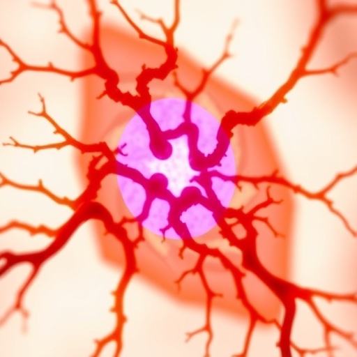
MINFLUX microscopy, an acronym for Minimal Fluorescence Photon Fluxes microscopy, introduces a paradigm shift in fluorescence imaging by leveraging a unique donut-shaped laser excitation pattern. This method excites individual fluorescent molecules with extreme precision, enabling scientists to track molecular events at resolutions finer than five nanometers and on temporal scales of milliseconds. This capability eclipses traditional confocal and super-resolution microscopes, offering unparalleled insights into the protein complexes that orchestrate neurotransmitter release and synaptic communication.
The necessity for extreme environmental control for MINFLUX’s operation cannot be overstated. The microscope demands installation within vibration-isolated and climate-controlled rooms because even minimal temperature fluctuations—as slight as two degrees—can introduce motion artifacts that degrade image fidelity. Such meticulous conditions highlight the intricacy and sensitivity of this instrument, underscoring the technical prowess required to harness its capabilities fully.
This substantial federal support hails from the National Institute of Standards and Technology (NIST) and was championed by Maryland Senators Angela Alsobrooks and Chris Van Hollen. Their advocacy culminated in the appropriation’s inclusion in the 2026 federal budget, a testimony to the strategic value of this technological advancement for national biomedical research priorities. The funding not only secures cutting-edge equipment but also facilitates the creation of a shared infrastructure designed to accelerate collaborative neuroscience research.
The microscopy facility is a cornerstone of the University of Maryland’s Medicine Institute for Neuroscience Discovery (UM-MIND) and will serve as a nexus for the MIND X-Change program. This consortium integrates multiple Maryland academic institutions to democratize access to state-of-the-art technologies, fostering an ecosystem aimed at unraveling the complexities of neurobiological disorders such as Alzheimer’s disease, profound autism, and treatment-resistant depression. By centralizing resources and expertise, UM-MIND is poised to catalyze discoveries that transcend individual laboratories.
Besides the MINFLUX system, the facility houses a lightsheet fluorescence microscope, capable of volumetric imaging of large brain tissue sections, perfectly complementing the ultra-high resolution approach of MINFLUX. The facility also plans to integrate a new confocal microscope to fill the intermediate imaging resolution niche. Confocal microscopy remains indispensable for visualizing cellular architecture in three dimensions, while lightsheet microscopy advances neural circuit mapping by preserving spatial relationships in large tissue volumes at high speed.
Technically, MINFLUX derives its extraordinary sensitivity from the spatially resolved excitation scheme, whereby fluorescent molecules are selectively activated in minuscule regions defined by the donut-shaped laser pattern. By positioning the excitation minimum at molecular coordinates, the technique localizes fluorescent emitters with unparalleled precision. This enables researchers to visualize intricate protein arrangements within synapses, investigate vesicular release mechanisms, and dissect the nanoscale choreography underpinning neuronal communication and plasticity.
The instrumentation’s ability to capture dynamic molecular processes in real-time opens new frontiers in understanding not only neurological functions but also pathological states, such as the metastatic behavior of cancer cells invading healthy brain tissues. In the pharmaceutical domain, MINFLUX holds promise in elucidating how therapeutic compounds modulate molecular assemblies and protein interactions, potentially accelerating drug discovery and optimization.
Given the sensitivity and resource-intensive nature of this technology, Dr. Tom Blanpied, Professor and Vice Chair of Pharmacology and Physiology, will oversee the microscopy core’s operations. Strategically located within UM-MIND’s facility in the Health Sciences Facility III (HSF-III), the core is designed as a collaborative hub that invites researchers and students alike to leverage these technologies to propel their investigations into uncharted cellular landscapes.
The broader vision of UM-MIND extends beyond microscopy to include a CRISPR-based genetic engineering core and the forthcoming AI and machine learning analytics core. The genetic engineering platform aims to facilitate advanced gene editing studies to demystify the neuronal signaling disruptions underlying disorders like autism and brain cancer. Meanwhile, the AI core will tackle the overwhelming data influx generated from imaging and behavioral experiments, refining data analysis and accelerating the extraction of meaningful biological insights.
A vivid example cited is the work of Dr. Margaret McCarthy, UM-MIND Director and UMSOM James and Carolyn Frenkil Dean’s Professor, whose studies on adolescent rat play behavior benefit from the AI-driven automated video analyses. This integration of nanoscopic imaging, genetic manipulation, and computational analysis epitomizes the multidisciplinary approach necessary to confront the complexity of the human brain and its diseases.
Dean Mark T. Gladwin of the University of Maryland School of Medicine articulates the significance of this investment, emphasizing that the availability of MINFLUX technology places the institution among a handful nationwide with nanoscale visualization capacity. The collective efforts of legislators and researchers encapsulate a strategic triumph poised to accelerate biomedical breakthroughs addressing some of medicine’s most intractable challenges—neurodegenerative diseases, addiction, and dementia.
Collectively, this advanced microscopy initiative at UM-MIND embodies a scientific renaissance where ultra-high-resolution visualization, genetic engineering precision, and artificial intelligence converge, propelling neuroscience from observing structures to understanding functions at the most fundamental molecular levels. As technology transcends previous limitations, the promise of deciphering the molecular underpinnings of brain function and dysfunction becomes an attainable frontier in biomedical science.
Subject of Research: Advanced fluorescence microscopy and neuroscience
Article Title: University of Maryland Secures $2.9 Million to Establish Ultra-High Resolution MINFLUX Microscopy Core for Cutting-Edge Neuroscience Discovery
News Publication Date: Not specified
Web References:
– https://www.medschool.umaryland.edu/um-mind/
– https://abberior.rocks/superresolution-confocal-systems/minflux/
– https://www.medschool.umaryland.edu/public-affairs/whats-the-buzz/december-2023/um-school-of-medicines-confocal-microscopy-facility-provides-zeiss-ls7-lightsheet-microscope-to-researchers-enhances-3d-visualization-of-biological-specimens.html
References: Not specified
Image Credits: University of Maryland School of Medicine
Keywords: MINFLUX microscopy, nanoscopy, fluorescence microscopy, cellular neuroscience, neurodegenerative diseases, synaptic imaging, CRISPR genetic engineering, artificial intelligence, drug discovery, advanced imaging technology
