Ultrasound can reliably track relative differences in cartilage thickness in the arthritic knee, but it consistently underestimates the true measurements that magnetic resonance imaging delivers, according to a new cross-sectional imaging study from India. The research, conducted at Mahatma Gandhi Medical College and Research Institute in Puducherry and published in the Egyptian Journal of Radiology and Nuclear Medicine, compared the two modalities side by side in 60 patients with knee osteoarthritis and found a striking gap between correlation and agreement: ultrasound followed MRI closely in its trends, yet its absolute numbers were systematically lower in every region of the medial femoral condyle examined.
The study focused on the medial femoral condyle, a key site of degeneration in knee osteoarthritis, a disease the authors describe as a leading cause of chronic pain and disability worldwide. The knee is the most commonly affected joint, with a reported community prevalence of 20 to 30 percent among Indian adults. Because conventional radiography cannot directly visualise cartilage, MRI has long served as the reference standard, offering high-contrast multiplanar images that allow accurate quantification of cartilage volume, thickness and defects. But MRI is costly, time-consuming and often impractical in routine or resource-limited care settings, which is precisely where ultrasound’s portability, real-time capability and low cost make it attractive as an alternative.
The research team enrolled 60 patients aged 40 or older with clinically diagnosed primary osteoarthritis who had been referred for MRI assessment. Participants presented with joint discomfort, crepitus or observable deformities, and the study excluded secondary osteoarthritis from systemic disorders, gout or rheumatoid arthritis, as well as patients with substantial prior knee trauma, previous joint surgery, or contraindications to either imaging modality. The cohort had a mean age of 56.23 years with a standard deviation of 6.99 years, and a mean body mass index of 27.6 kilograms per square metre. Women made up a slight majority at 53.3 percent. Kellgren–Lawrence grading showed a cohort skewed towards early disease: Grade 1 in 46.7 percent of participants, Grade 2 in 26.7 percent, Grade 3 in 18.3 percent and Grade 4 in 8.3 percent.
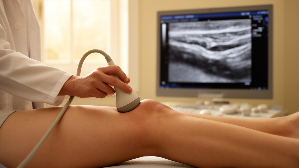
Ultrasound examinations of the medial femoral articular cartilage of the left knee were performed with a 10 MHz linear transducer while the knee was flexed at 90 degrees. Cartilage thickness was measured as the perpendicular distance between the superficial and deep hyperechoic interfaces. A single trained radiologist carried out all ultrasound scans without knowledge of the corresponding MRI measurements, a design choice intended to minimise inter-observer variability. The left knee was assessed in all subjects for protocol uniformity, including in cases of bilateral disease. The anterior, middle and posterior sub-regions of the medial femoral condyle were defined on ultrasound using a cylinder-fitting technique aligned with the femoral condylar anatomy, and the corresponding sub-regions were located on the identical mid-condylar sagittal MRI slice using the intercondylar notch and posterior femoral cortex as stable bony references, ensuring that both modalities assessed anatomically comparable segments. MRI was performed on a 1.5 Tesla system with a dedicated knee coil, with thickness measured on the workstation at the anterior, middle and posterior regions of the sagittal slice representing the midpoint of the medial femoral condyle.
The results revealed a consistent pattern. MRI recorded greater mean medial femoral cartilage thickness than ultrasound in every region: 1.35 millimetres anteriorly, 1.66 millimetres in the middle and 2.05 millimetres posteriorly, compared with 0.91, 0.97 and 0.99 millimetres respectively on ultrasound. The discrepancy was most pronounced in the posterior region. Strong positive correlations between the modalities were observed across all three regions, with Pearson correlation coefficients ranging from 0.767 to 0.904 and p values below 0.001. Yet intraclass correlation coefficient analysis, using a two-way random-effects model for absolute agreement, told a different story: only moderate agreement in the anterior region (ICC 0.541) and poor agreement in the middle (ICC 0.416) and posterior (ICC 0.264) regions. In other words, ultrasound reflects relative trends in cartilage thickness among individuals but has limited absolute concordance with MRI.
Bland–Altman analysis confirmed the systematic underestimation. Mean bias was 0.44 millimetres anteriorly, with 95 percent limits of agreement from −0.38 to 1.25 millimetres, and 1.06 millimetres posteriorly, with limits of agreement from −0.10 to 2.22 millimetres. The analysis also revealed a proportionate bias, with the discrepancy between modalities increasing as cartilage thickness increased, particularly in the middle and posterior regions. The authors note that a 1 millimetre underestimation in the posterior region accounts for nearly half of the total cartilage thickness measured by MRI there, underscoring the need for clinicians to apply modality-specific bias correction when interpreting ultrasound measurements. As an exploratory step towards correction, the team graphed the three regional mean biases against their respective regional mean thicknesses and observed a positive, steadily increasing relationship, with an approximate slope of 1.6 millimetres of bias for each millimetre rise in mean thickness. The authors caution that this exploratory analysis rests on only three regional aggregate points rather than individual patient-level pairs, and that a formal linear regression of per-patient differences against per-patient means is needed to statistically validate the trend.
The discussion offers several explanations for the systematic gap. Transducer pressure, the anisotropic properties of cartilage, and the technical challenge of reliably identifying the cartilage–bone interface on ultrasound all contribute. The authors also point to their equipment: the 10 MHz linear transducer used in the study, while sufficient for general knee musculoskeletal imaging, has inferior axial resolution compared with the 12 to 18 MHz probes increasingly used for high-resolution superficial cartilage morphometry. This reduced axial resolution likely enlarged the perceived cartilage–bone interface and contributed to the systematic underestimation, especially posteriorly, where beam obliquity exacerbates the problem. The poor posterior ICC likely also reflects the difficulty of maintaining a perpendicular beam against the convex posterior condyle, producing refraction artefacts and an indistinct bone–cartilage interface, a limitation that does not affect MRI. The authors call for future research with higher-frequency transducers to determine whether resolution, rather than condylar curvature alone, accounts for the posterior underestimation.
On demographics, age was significantly associated with cartilage thinning in the middle and posterior regions on MRI and in the middle region on ultrasound, with p values below 0.05, a finding consistent with the known pathophysiology of osteoarthritis, in which biochemical and structural degeneration progresses over time. No significant associations were identified between cartilage thickness and gender or body mass index, which the authors attribute to the homogeneous nature of a symptomatic osteoarthritis population or region-specific measurement effects. As a preliminary corrective measure, the team formulated region-specific linear prediction equations based on the documented regional averages, standard deviations and correlation coefficients: anterior, MRI approximately equals 0.87 times ultrasound plus 0.56 millimetres; middle, 1.48 times ultrasound plus 0.23 millimetres; posterior, 1.72 times ultrasound plus 0.34 millimetres. The authors stress that these equations serve as examples rather than conclusive results, because they derive from collective regional data instead of patient-specific regression, and formal derivation and cross-validation using individual paired measurements is advisable before any clinical use.
The study’s authors conclude that, while MRI remains the gold standard for precise cartilage morphometry, ultrasound shows promise as a complementary tool. Given its strong correlation but limited agreement with MRI, they position ultrasound as suitable for screening and longitudinal monitoring of relative cartilage changes, particularly in resource-limited settings, but not as a replacement for MRI in definitive quantitative cartilage evaluation. The findings align with previous work, including studies by Schmitz and colleagues, who reported ultrasound measurements 1.9 to 2.8 millimetres lower than MRI, and by Yoon and colleagues, who reported significant correlations in specific femoral regions, as well as work by Naredo and colleagues showing that ultrasound measurements correlate better in medial condyle regions than in deeper posterior zones.
The authors are candid about the limitations. The design was cross-sectional and single-centre, the sample size was 60, and ultrasound is operator dependent. The cohort’s bias towards early-stage disease, with Kellgren–Lawrence Grades 1 and 2 accounting for 73.4 percent of individuals, prevented a statistically sound stratified analysis of ultrasound–MRI concordance between early and advanced-stage osteoarthritis. Assessment was limited to the left knee, excluding bilateral or right-knee disease, and no formally quantified inter- or intra-observer reliability assessment for ultrasound was performed, since repeat measurements by a second examiner were not conducted. Because cartilage thickness is a continuous variable, sensitivity, specificity and ROC analyses were not applied. Future studies, the authors suggest, should include larger multicentre cohorts with longitudinal follow-up, standardised ultrasound scanning protocols, structured operator training, documentation of subgroup concordance by Kellgren–Lawrence grade, assessment of both knees, and a focused investigation of inter- and intra-observer repeatability within a patient subset. Until then, the message for clinicians is measured: ultrasound offers an accessible, economical window on cartilage trends in the osteoarthritic knee, but its absolute numbers must be interpreted with the modality’s known bias in mind.
Subject of Research: Correlation and agreement between ultrasound and MRI in measuring medial femoral condyle cartilage thickness in knee osteoarthritis
Article Title: Utility of ultrasound as a diagnostic tool for measuring knee cartilage thickness in patients with osteoarthritis: correlation with MRI
Article References: Purushotham, K., Kumar, K., Prabakaran, T., Lokesh Kumar, T., & Manesha, P. (2026). Utility of ultrasound as a diagnostic tool for measuring knee cartilage thickness in patients with osteoarthritis: correlation with MRI. Egyptian Journal of Radiology and Nuclear Medicine, 57(1), Article 226. https://doi.org/10.1186/s43055-026-01868-z
Image Credits: AI Generated
DOI: 10.1186/s43055-026-01868-z
Keywords: ultrasound, MRI, knee osteoarthritis, cartilage thickness, medial femoral condyle, Bland-Altman analysis, intraclass correlation coefficient, medical imaging, radiology, cartilage morphometry, diagnostic imaging, resource-limited settings
Cite Scienmag News
Ophelia Keating. (October 28, 2026). Ultrasound tracks knee cartilage loss but falls short of MRI precision. Scienmag. https://scienmag.com/ultrasound-tracks-knee-cartilage-loss-but-falls-short-of-mri-precision/
Ophelia Keating. "Ultrasound tracks knee cartilage loss but falls short of MRI precision." Scienmag, 28 October 2026, https://scienmag.com/ultrasound-tracks-knee-cartilage-loss-but-falls-short-of-mri-precision/. Accessed 28 October 2026.
Ophelia Keating. "Ultrasound tracks knee cartilage loss but falls short of MRI precision." Scienmag. October 28, 2026. https://scienmag.com/ultrasound-tracks-knee-cartilage-loss-but-falls-short-of-mri-precision/