


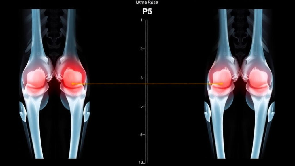
A small pad of fat tucked behind the kneecap has become the unlikely star of a new study that could change how doctors evaluate one of the most common complaints in sports medicine: anterior knee pain. Researchers in Turkey report that a technique called shear wave elastography, which measures how stiff living tissue is in real time, can detect edema in the suprapatellar fat pad with remarkable accuracy, distinguishing affected knees from healthy ones with an area under the curve of 0.949. The finding, published in BMC Medical Imaging, is the first time this ultrasound-based stiffness mapping method has been applied to the suprapatellar fat pad, and it opens a tantalizing possibility: that a quick, radiation-free, and relatively cheap bedside scan might one day flag a problem that currently requires a costly magnetic resonance imaging session to confirm.
The suprapatellar fat pad is a triangular wedge of fat lying directly behind the quadriceps tendon, just above the patella, inside the joint capsule region of the knee. For years it was treated as anatomical filler, a passive cushion between tendon and bone. That view has shifted. Magnetic resonance imaging studies have shown that edema, an accumulation of fluid within the pad, is a frequent finding, and clinicians now associate it with suprapatellar fat pad impingement syndrome, a condition in which the pad gets pinched between the quadriceps tendon and the femur during knee extension. The result is often anterior knee pain, a symptom that plagues runners, cyclists, office workers who sit all day, and a large share of patients who walk into orthopedic and rheumatology clinics. The problem is that MRI, the gold standard for spotting this edema, is expensive, time-consuming, and unavailable to many patients at the point of care.
Shear wave elastography offers a fundamentally different way to interrogate tissue. Instead of relying on the subjective eyeballing of grayscale ultrasound images, SWE generates precisely controlled mechanical vibrations deep within the tissue using the acoustic radiation force of focused ultrasound pulses. These vibrations produce shear waves, transverse ripples that propagate sideways through the tissue much like ripples spreading across a pond. The crucial physics is that the speed of these shear waves depends directly on the elasticity of the medium: stiffer tissue, such as fibrotic liver or inflamed fat, transmits the waves faster. By tracking the wave propagation with ultrafast ultrasound imaging and applying a Young’s modulus conversion, the system outputs quantitative stiffness values in kilopascals. The technique has already earned clinical acceptance in liver fibrosis staging, breast lesion characterization, and thyroid and musculoskeletal applications, but no one had systematically measured the suprapatellar fat pad with it until now.
The research team, led by Muhammed Erkam Çeker of Tokat State Hospital together with Fatih Çelikyay of Sincan Training and Research Hospital in Ankara and Sevgi Yılmaz Sağlam of Ordu University Hospital, designed a prospective case-control study to test whether edema visible on MRI corresponds to measurable changes in tissue stiffness. They enrolled 35 adult patients whose MRI examinations had confirmed suprapatellar fat pad edema, along with 31 control participants whose knee MRIs, obtained for unrelated reasons, showed no edema. Crucially, the elastography examination was performed within seven days of the MRI, minimizing the chance that the tissue state changed between the two imaging modalities. The radiologist performing the ultrasound measurements was blinded to which group each participant belonged to, a safeguard against unconscious bias in where to place measurement regions or how to interpret borderline readings.
The measurement protocol was deliberately systematic. Because the suprapatellar fat pad is triangular, the team sampled stiffness at three predefined corner regions: the superior apex, designated S1; the anteroinferior corner, S2; and the posteroinferior corner, S3. In addition, they manually traced the entire fat pad in a single longitudinal ultrasound plane and recorded a whole-area stiffness value, designated SA. This dual approach, point sampling plus area averaging, was intended to capture both localized and global changes in the pad’s mechanical properties. All values were expressed in kilopascals, and statistical comparisons between the edema and control groups used the independent-samples t-test or the Mann-Whitney U test as appropriate, with Bonferroni correction applied across the four parameters to guard against false positives from multiple testing.
The results were striking. Mean whole-area stiffness in the edema group was 26.14 plus or minus 8.69 kilopascals, roughly double the 13.14 plus or minus 3.04 kilopascals recorded in controls, a difference that was highly significant with a p-value below 0.001. The corner measurements told the same story: stiffness at all three sampled regions was significantly elevated in the edema group, and every one of those differences survived the stringent Bonferroni correction. In other words, the effect was not an artifact of cherry-picking the most favorable measurement site; the entire fat pad had become measurably harder in knees with MRI-confirmed edema. From a biomechanical standpoint, this makes intuitive sense, since fluid infiltration and inflammatory change within adipose tissue would be expected to alter its viscoelastic behavior, and shear wave speed is exquisitely sensitive to exactly those changes.
To quantify how well stiffness could discriminate between the two groups, the researchers performed receiver operating characteristic analysis, the standard method for evaluating diagnostic tests. The area under the curve, which ranges from 0.5, equivalent to a coin flip, to 1.0, representing perfect discrimination, reached 0.949 for the whole-area measurement and 0.916 for the posteroinferior corner S3. Values above 0.9 are conventionally considered excellent, placing this simple ultrasound measurement in the same diagnostic performance tier as many established imaging tests. The authors derived cutoff values within their cohort, though they were careful to note that these thresholds were generated and evaluated in the same dataset, a practice that tends to produce optimistic estimates and requires validation in independent populations before any clinical adoption.
Two subgroup findings, or rather the absence of findings, deserve attention. Stiffness did not differ significantly between patients who reported anterior knee pain and those who did not, nor between cases classified as mild edema and those classified as advanced. The authors report p-values above 0.05 for all such comparisons, but they also acknowledge that these subgroup analyses had limited statistical power, meaning the small sample sizes make it impossible to conclude that no relationship exists. This nuance matters for interpretation: the study establishes that edema and stiffness go together, but whether stiffness tracks symptom severity or edema grade remains an open question that only larger cohorts can answer.
The authors are appropriately candid about the limitations of their work, a transparency that strengthens rather than weakens the study. The control group, recruited from patients scanned for unrelated indications, was not clinically representative of people who present with knee complaints, so the comparison, while clean, does not replicate the messy diagnostic reality of a pain clinic. The cutoff values were derived and tested in the same cohort rather than in a separate validation set. And a single operator performed all elastography measurements, with no reproducibility assessment, leaving open the question of how much the results would vary between different sonographers, machines, or institutions. The study was approved by the Ethics Committee of Tokat Gaziosmanpaşa University in May 2022, conducted under the Declaration of Helsinki, and all participants gave written informed consent. No funding was received, and the authors declare no competing interests.
Even with those caveats, the implications are considerable. If future studies with larger, clinically representative cohorts and standardized, reproducible protocols confirm these results, shear wave elastography could become a rapid first-line tool for evaluating suspected suprapatellar fat pad impingement, reserving MRI for complex or ambiguous cases. That would mean faster diagnoses, lower costs, and no ionizing radiation or claustrophobic scanner tubes for millions of knee pain sufferers. It would also add a new chapter to the growing recognition that fat pads around joints, long dismissed as inert packing material, are dynamic structures whose mechanical properties carry real diagnostic information. For now, the message from Tokat, Ankara, and Ordu is a proof of concept: a common MRI finding leaves a measurable mechanical fingerprint that a handheld ultrasound probe can read in kilopascals, and that fingerprint is nearly twice as stiff in affected knees. The next step, validating it in the clinic, is one the field will be watching closely.
Subject of Research: Shear wave elastography measurement of suprapatellar fat pad stiffness in patients with MRI-confirmed edema
Article Title: Shear wave elastography of the suprapatellar fat pad in patients with MRI-confirmed edema: a case-control study
Article References: Çeker, M. E., Çelikyay, F., & Sağlam, S. Y. (2026). Shear wave elastography of the suprapatellar fat pad in patients with MRI-confirmed edema: a case-control study. BMC Medical Imaging. https://doi.org/10.1186/s12880-026-02906-y
Image Credits: AI Generated
DOI: 10.1186/s12880-026-02906-y
Keywords: shear wave elastography, suprapatellar fat pad, knee edema, MRI, anterior knee pain, impingement syndrome, musculoskeletal ultrasound, tissue stiffness, case-control study, radiology, diagnostic imaging, orthopedics
Cite Scienmag News
Ophelia Keating. (October 10, 2026). Ultrasound Stiffness Test Detects Knee Fat Pad Edema With Striking Accuracy. Scienmag. https://scienmag.com/ultrasound-stiffness-test-detects-knee-fat-pad-edema-with-striking-accuracy/
Ophelia Keating. "Ultrasound Stiffness Test Detects Knee Fat Pad Edema With Striking Accuracy." Scienmag, 10 October 2026, https://scienmag.com/ultrasound-stiffness-test-detects-knee-fat-pad-edema-with-striking-accuracy/. Accessed 10 October 2026.
Ophelia Keating. "Ultrasound Stiffness Test Detects Knee Fat Pad Edema With Striking Accuracy." Scienmag. October 10, 2026. https://scienmag.com/ultrasound-stiffness-test-detects-knee-fat-pad-edema-with-striking-accuracy/


