Morphea, also known as localized scleroderma, is an autoimmune connective tissue disorder in which the immune system attacks the skin and sometimes the tissue beneath it, driving inflammation and progressive fibrosis that can leave permanent scars, disfigurement, and joint contractures. Unlike systemic sclerosis, morphea is confined to the skin and subcutaneous structures, but the burden it places on patients, particularly children, can be substantial. For decades, dermatologists have relied on clinical scoring systems such as the Localized Scleroderma Activity Index (LoSAI) and the Localized Scleroderma Damage Index (LoSDI), along with physician global assessments, to judge whether a lesion is active and whether treatment is working. The problem is that these tools are inherently subjective. Palpation of skin thickening varies from one examiner to another, erythema can be difficult to discern in darker skin tones, and the difference between active inflammation and residual damage is often invisible to the naked eye. A new prospective study published in the Archives of Dermatological Research suggests that high-frequency ultrasound, color Doppler imaging, and shear-wave elastography could give clinicians the objective, measurable biomarkers they have long lacked for tracking treatment response in this stubborn disease.
The research, led by Faezeh Khorasanizadeh of the Department of Radiology at Razi Hospital, Tehran University of Medical Sciences, together with colleagues in dermatology, epidemiology, and biostatistics, and in collaboration with Ximena Wortsman of the Universidad de Chile, a pioneer in cutaneous ultrasound, enrolled eighteen patients with newly biopsy-confirmed morphea. Every participant had at least one clinically active plaque at baseline. The investigators deliberately designed the protocol to be practical: rather than imaging every lesion, they selected the most clinically probable active plaque in each patient and compared it with adjacent or contralateral normal skin serving as an internal control. Each patient then underwent grayscale ultrasound, color Doppler ultrasound, and shear-wave elastography at the start of the study and again after six months of treatment, alongside standardized clinical scoring. The study received ethical approval from the institutional review board of Tehran University of Medical Sciences and was conducted in accordance with the Declaration of Helsinki, with written informed consent obtained from all participants and guardians of minors.
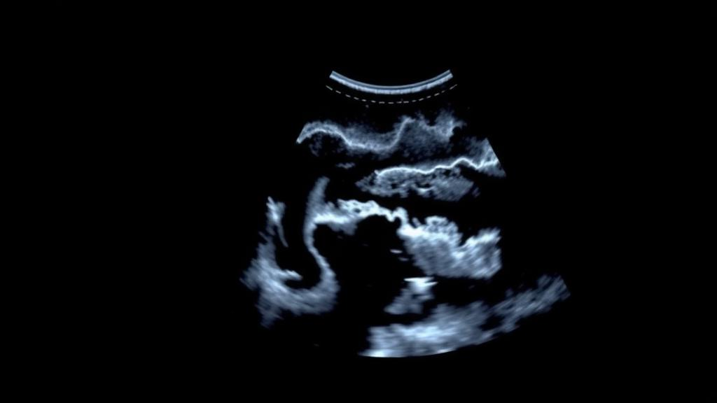
The technical logic behind the imaging approach is worth unpacking. High-frequency grayscale ultrasound can resolve the individual layers of the skin with millimeter precision, allowing radiologists to measure dermal thickness, the hallmark of fibrotic thickening in morphea, and hypodermal thickness, which reflects the fat layer beneath. In active lesions, inflammation and edema typically thicken the dermis and can alter the echotexture of the subcutis. Color Doppler ultrasound adds a functional dimension by detecting blood flow; increased vascularity within and beneath a plaque is considered a marker of subclinical inflammatory activity that may persist even when the skin surface appears quiescent. Shear-wave elastography, the most technically sophisticated of the three, works by generating shear waves, a form of transverse mechanical vibration, within the tissue and measuring the speed at which they propagate. Because stiffer tissue transmits shear waves faster than soft tissue, the technique yields a quantitative estimate of tissue elasticity in kilopascals, turning the subjective sensation of hardened skin into a reproducible number.
After six months of follow-up, the results told a clear and clinically meaningful story. Dermal thickness, measured ultrasonographically, decreased significantly alongside a significant fall in mean clinical scores, with p values below 0.05. The LoSAI activity score dropped from a mean of 4.28 plus or minus 3.28 at baseline to 1.44 plus or minus 1.75 after treatment, while the LoSDI damage score declined from 11.39 plus or minus 7.38 to 9.56 plus or minus 6.77. Hypodermal thickness also decreased and skin elasticity increased slightly over the six months, but neither of these secondary changes reached statistical significance. The pattern makes biological sense: inflammation-driven dermal thickening responds to immunosuppressive therapy within months, whereas fibrotic remodeling of the subcutis and restoration of elastic properties are slower processes that may lag behind clinical improvement or, in the case of established damage, never fully reverse.
Perhaps the most intriguing findings emerged from the correlation analyses. Hypodermal thickness correlated negatively with the Physician Global Assessment of Damage, with a Spearman rho of -0.410 and a p value of 0.027, and positively with the LoSAI activity index, with rho of 0.513 and p of 0.004. Hypodermal thickness was also positively correlated with the Physician Global Assessment of Activity before treatment began. After therapy, hypodermal stiffness measured by elastography correlated positively with the LoSDI damage index. Taken together, these relationships suggest that the subcutaneous fat layer, often overlooked in favor of the dermis, carries real diagnostic information: a thicker hypodermis tracks with active disease, while stiffer subcutaneous tissue tracks with accumulated damage. This positions elastography not merely as a research curiosity but as a potential bridge between the activity and damage domains that clinicians must distinguish when deciding whether to escalate, continue, or taper therapy.
Not every result favored the new technology, and the authors were candid about the limitations. When radiologists and dermatologists independently rated disease activity using shear-wave elastography and color Doppler, the inter-rater agreement was poor, with a kappa of just 0.053, barely above chance. This discordance likely reflects the steep learning curve of cutaneous ultrasound, differences in probe positioning and pressure, and the absence of universally standardized scoring protocols across specialties. It is a sobering reminder that a measurement technique is only as objective as the protocol governing its use. The authors also emphasized that their cohort was small, with only eighteen patients, all of whom had plaque-type or linear morphea, the most common subtypes. Generalizing to rarer variants such as generalized, deep, or pansclerotic morphea will require larger and more diverse cohorts, and the six-month follow-up window may be too short to capture slower changes in elasticity and subcutaneous architecture.
Why does an objective biomarker matter so much for morphea specifically? Current treatment typically involves systemic corticosteroids combined with methotrexate or, increasingly, other immunomodulators, a regimen that carries real toxicity and demands months of commitment. Because clinical scores are subjective and damage can masquerade as activity, physicians risk both overtreating patients whose disease has already burned out and undertreating those with smoldering subclinical inflammation that continues to cause irreversible fibrosis. Ultrasound-based measures could change this calculus in several ways: by providing early, quantitative evidence of response within weeks rather than months; by identifying subclinical activity that warrants continued therapy even when the skin looks better; and by serving as surrogate endpoints in clinical trials, where the rarity of morphea has long made adequately powered studies difficult. Prior work by Wortsman and colleagues on the Ultrasound Morphea Activity Scoring system, and by other groups applying shear-wave elastography in both localized and systemic scleroderma, has laid the groundwork, but this study is among the first to track patients longitudinally through treatment with a multimodal protocol.
The study also fits into a broader movement in dermatology toward quantitative, imaging-based assessment of skin disease. Thermal imaging has been explored for detecting inflammation in localized scleroderma, dermoscopy offers clues to disease stage, and 50-megahertz ultrasound has documented improvement after ultraviolet A1 phototherapy. In systemic sclerosis, elastography is being evaluated as a measure of skin strain and fibrosis severity, raising the possibility of a shared imaging vocabulary across the sclerosing diseases. A recent consensus statement on the diagnosis and treatment of sclerosing skin diseases underscores how much remains unsettled in monitoring these conditions, and the authors of the current study position their work as a preliminary but prospective step toward filling that gap with tools that are already available in most radiology departments.
For now, the take-home message is measured but promising. In this preliminary prospective cohort, dermal thickness measured with high-frequency ultrasound emerged as the strongest candidate for an objective marker of treatment response in morphea, while color Doppler and shear-wave elastography showed potential as adjunctive tools whose clinical utility remains exploratory. The authors themselves stress that validation in larger cohorts is essential before these techniques enter routine practice, and the poor inter-rater agreement highlights the need for standardized acquisition and scoring protocols, ideally with dedicated training for both radiologists and dermatologists. Still, the direction of travel is unmistakable. If subsequent studies confirm these findings, the era of guessing whether a morphea plaque is truly quiet could give way to an era in which a handheld probe, a quantitative elasticity map, and a few millimeters of measured dermis tell clinicians exactly what the immune system is doing beneath the skin, and whether the treatment they have chosen is actually working.
Subject of Research: Ultrasound and shear-wave elastography for monitoring treatment response in morphea
Article Title: Ultrasound and shear-wave elastography as a tool for evaluating response to treatment of morphea: a preliminary report of a prospective study
Article References: Khorasanizadeh, F., Sharifi, M. A., Vahabi, S. M., Ansari, M. S., Ghandi, N., Hosseini, F., Etesami, I., & Wortsman, X. (2026). Ultrasound and shear-wave elastography as a tool for evaluating response to treatment of morphea: a preliminary report of a prospective study. Archives of Dermatological Research, 318(1), Article 457. https://doi.org/10.1007/s00403-026-04919-7
Image Credits: AI Generated
DOI: 10.1007/s00403-026-04919-7
Keywords: morphea, localized scleroderma, ultrasound, shear-wave elastography, color Doppler, dermal thickness, LoSAI, LoSDI, treatment response, biomarkers, dermatology, skin fibrosis
Cite Scienmag News
Ophelia Keating. (October 7, 2026). Ultrasound Offers an Objective Window Into Morphea Treatment Response. Scienmag. https://scienmag.com/ultrasound-offers-an-objective-window-into-morphea-treatment-response/
Ophelia Keating. "Ultrasound Offers an Objective Window Into Morphea Treatment Response." Scienmag, 7 October 2026, https://scienmag.com/ultrasound-offers-an-objective-window-into-morphea-treatment-response/. Accessed 7 October 2026.
Ophelia Keating. "Ultrasound Offers an Objective Window Into Morphea Treatment Response." Scienmag. October 7, 2026. https://scienmag.com/ultrasound-offers-an-objective-window-into-morphea-treatment-response/