


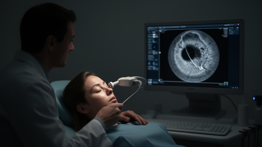
A 72-year-old woman whose persistent eye redness, pain, and progressive vision loss had been treated as uveitis turned out to have a rare, hidden eye cancer that standard imaging could not see. The case, published as an open-access case report in Discover Oncology on 27 October 2026, describes an amelanotic ciliary body melanoma that remained undetected on scanning laser ophthalmoscopy, anterior segment optical coherence tomography, B-scan ultrasonography, and orbital magnetic resonance imaging, and was only revealed by ultrasound biomicroscopy.
Amelanotic ciliary body melanoma is a rare and diagnostically challenging intraocular malignancy. Because it lacks the pigment that makes most uveal melanomas visible, it can masquerade as anterior segment inflammation and evade conventional imaging, as this case illustrates. On examination, the patient had iris neovascularization, focal iris elevation, localized obstruction of the drainage angle, and an intraocular pressure of 29 mmHg — signs of serious intraocular disease, yet no definite mass could be identified on any of the conventional studies.
Local excision of the lesion confirmed amelanotic melanoma composed predominantly of small round-to-oval cells, with a Ki-67 proliferation index of approximately 30%.
The clinical course then took a difficult turn. Postoperative hyphema — bleeding into the front chamber of the eye — and refractory ocular hypertension could not be controlled, and the eye ultimately required enucleation. Final pathological examination of the enucleated specimen showed residual melanoma with predominantly epithelioid morphology involving the ciliary body and adjacent choroid, with invasion of the retina and a markedly higher Ki-67 index of approximately 60%.
Immunohistochemistry revealed striking discordance between the two specimens. SOX10 and PRAME were positive in both the initial excision and the enucleation specimen, whereas the expression of S100, Melan-A, and HMB45 differed between them. The authors highlight this marked pathological discordance between sequential specimens as evidence that intratumoral heterogeneity must be considered in ciliary body melanoma, alongside careful clinicopathological correlation.
At 12 months of follow-up, no orbital recurrence or distant metastasis had been detected. The report’s central conclusions are twofold: UBM enabled detection of an otherwise occult amelanotic ciliary body melanoma after nondiagnostic conventional imaging, and the confirmed retinal invasion together with the changing histopathological profile underscores both the diagnostic value of UBM in unexplained anterior segment disease and the biological variability these tumors can display. As a single case report, the findings describe one patient’s experience rather than a systematic evaluation of UBM’s diagnostic performance, but they illustrate why clinicians may need to look beyond standard imaging when inflammatory masquerade syndromes fail to respond to treatment.
Subject of Research: Detection of an occult amelanotic ciliary body melanoma by ultrasound biomicroscopy after nondiagnostic conventional imaging
Article Title: Occult amelanotic ciliary body melanoma with retinal invasion detected by ultrasound biomicroscopy after nondiagnostic conventional imaging: a case report
Article References: Shen, Y., Huang, R., Jiang, A., Ji, P., & Yan, M. (2026). Occult amelanotic ciliary body melanoma with retinal invasion detected by ultrasound biomicroscopy after nondiagnostic conventional imaging: a case report. Discover Oncology. https://doi.org/10.1007/s12672-026-06204-5
Image Credits: AI Generated
DOI: 10.1007/s12672-026-06204-5
Keywords: ciliary body melanoma, amelanotic melanoma, ultrasound biomicroscopy, ocular oncology, retinal invasion, intratumoral heterogeneity, Ki-67 index, enucleation, uveitis masquerade, case report, Occult, amelanotic
Cite Scienmag News
Nathaniel Bowman. (October 27, 2026). Ultrasound Biomicroscopy Detects Occult Ciliary Body Melanoma Missed by Conventional Imaging. Scienmag. https://scienmag.com/ultrasound-biomicroscopy-detects-occult-ciliary-body-melanoma-missed-by-conventional-imaging/
Nathaniel Bowman. "Ultrasound Biomicroscopy Detects Occult Ciliary Body Melanoma Missed by Conventional Imaging." Scienmag, 27 October 2026, https://scienmag.com/ultrasound-biomicroscopy-detects-occult-ciliary-body-melanoma-missed-by-conventional-imaging/. Accessed 27 October 2026.
Nathaniel Bowman. "Ultrasound Biomicroscopy Detects Occult Ciliary Body Melanoma Missed by Conventional Imaging." Scienmag. October 27, 2026. https://scienmag.com/ultrasound-biomicroscopy-detects-occult-ciliary-body-melanoma-missed-by-conventional-imaging/


