


A 64-year-old man presented with chest pain and an inferior ST-elevation myocardial infarction. His cardiovascular profile was already heavily loaded: he was an ex-smoker with hypertension, dyslipidemia, and atrial fibrillation, and five years earlier he had undergone percutaneous coronary intervention of the proximal right coronary artery, where a 2.75 × 8 mm drug-eluting stent had been implanted. Despite optimal medical treatment, including dabigatran, his left ventricular ejection fraction was preserved at 60%, and rosuvastatin 40 mg/day kept his low-density lipoprotein cholesterol at 63 mg/dL.
What clinicians found when they imaged the culprit artery turned out to be unprecedented in the published literature. Right coronary artery angiography revealed severely calcified diffuse disease, with consecutive intermediate stenoses in the proximal and mid segments and a significant stenosis in the mid-distal segment. The calcification was so pronounced that it hindered visualization of the previously implanted stent, and the diffuse pattern with multiple stenoses made angiography alone insufficient.
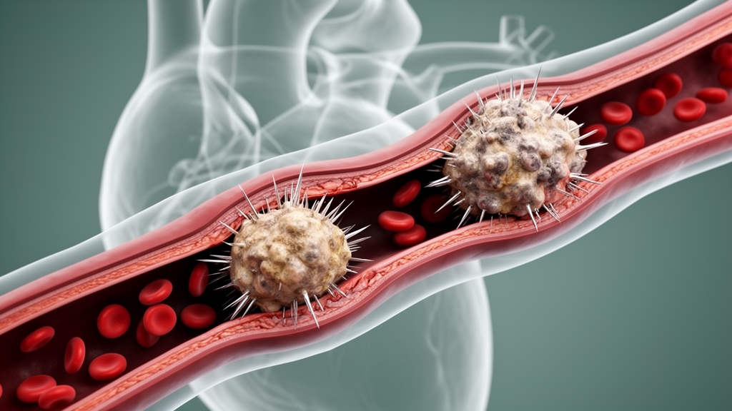
The team therefore turned to optical coherence tomography, which delivered a striking answer: two eruptive calcified nodules, one in the proximal segment just distal to the old stent and a second in the mid-distal segment. To the best of the authors' knowledge, this is the first reported case of two eruptive calcified nodules located within the same artery in a patient presenting with a myocardial infarction. Eruptive calcified nodules are rare but consequential.
They represent the least frequent morphology, roughly 5% of cases, causing thrombotic acute coronary syndrome, and they are characterized by a disrupted fibrous cap, often accompanied by an overlying fibrin or platelet thrombus that gives the lesion an irregular surface. These lesions are almost universally deformable because of their composition of fragmented necrotic core calcification. The finding of two such nodules in one artery suggests that calcified nodule destabilization may occur concurrently, in a manner analogous to plaque rupture, pointing toward a shared underlying pathophysiological process that could involve increased vascular inflammatory activity.
The authors are careful to note, however, that this interpretation remains speculative and cannot be established from a single case. Which nodule triggered the heart attack? The distal calcified nodule was considered the presumed culprit lesion based on the angiographic findings and the associated thrombotic appearance.
The proximal nodule was regarded as a non-culprit lesion, although its potential contribution to the acute coronary event cannot be completely excluded. Calcified nodules are a challenging lesion subset associated with decreased procedural success and worse clinical outcomes, and the optimal treatment strategy has yet to be determined. In this case, the presence of thick circumferential calcium along with deep medial calcification led the team to use coronary intravascular lithotripsy (Shockwave Medical, Santa Clara, CA), with the aim of facilitating adequate lesion preparation and stent expansion.
Sequential pulse treatments were applied with 3.0 × 12 mm and 3.5 × 12 mm balloons, working from the mid-distal to the proximal right coronary artery. Importantly, the clinicians treated the entire calcified segment rather than only the two visually apparent nodules, given the diffuse calcified disease extending beyond them. After dilatation with non-compliant balloons, two overlapping drug-eluting stents were implanted, measuring 3.0 × 48 mm and 4.0 × 32 mm.
That decision reflects accumulating evidence about how these lesions behave during intervention. Although optimal disruption of calcium with intravascular lithotripsy is best achieved when the coronary calcification is circumferential, a recent optical coherence tomography analysis revealed that its application was safe and highly effective in treating calcified nodules, leading to adequate stent expansion. That same analysis uncovered a subtlety with direct implications for stenting.
In a pooled analysis, the baseline minimal lumen area coincided with the location of the site of maximum calcified nodule in only 51.7% of cases, and in 57.7% of cases the minimal stent area after intervention was located outside the maximum calcified nodule site. In other words, the narrowest point before treatment and the weakest stent expansion after treatment frequently do not sit at the most prominent nodule. The practical conclusion is that treatment approaches should target not only the calcified nodules themselves but also the entire calcified lesion, exactly as was done here.
The clinical outcome so far is reassuring. The patient remains asymptomatic and event-free at four-year follow-up, a durable result in a lesion subset known for procedural difficulty and adverse outcomes. The case also demonstrates a concrete diagnostic capability: optical coherence tomography can identify multiple eruptive calcified nodules in the same coronary artery, and that identification can influence treatment planning, from the choice of calcium-modification technique to the length of the stented segment.
Whether simultaneous nodule destabilization truly mirrors the multifocal behavior of plaque rupture, and whether a shared inflammatory driver underlies such presentations, will require systematic study. What the case establishes is that when diffuse calcified coronary disease presents as an acute syndrome, more than one eruptive nodule may lurk within the same artery, and imaging that resolves the artery wall can reveal them both before the stents go in.
Subject of Research: Cardiology
Article Title: Two Eruptive Calcified Nodules in the Same Artery
Article References: Andreou, I., & Sabaté, M. (2026). Two Eruptive Calcified Nodules in the Same Artery. Clinical Case Reports, 14(11), Article e73648. https://doi.org/10.1002/ccr3.73648
Image Credits: AI Generated
DOI: 10.1002/ccr3.73648
Keywords: Eruptive, Calcified, Nodules, Same, Artery, scientific research
Cite Scienmag News
Ophelia Keating. (October 26, 2026). Two Eruptive Calcified Nodules Found in the Same Coronary Artery. Scienmag. https://scienmag.com/two-eruptive-calcified-nodules-found-in-the-same-coronary-artery/
Ophelia Keating. "Two Eruptive Calcified Nodules Found in the Same Coronary Artery." Scienmag, 26 October 2026, https://scienmag.com/two-eruptive-calcified-nodules-found-in-the-same-coronary-artery/. Accessed 26 October 2026.
Ophelia Keating. "Two Eruptive Calcified Nodules Found in the Same Coronary Artery." Scienmag. October 26, 2026. https://scienmag.com/two-eruptive-calcified-nodules-found-in-the-same-coronary-artery/


