


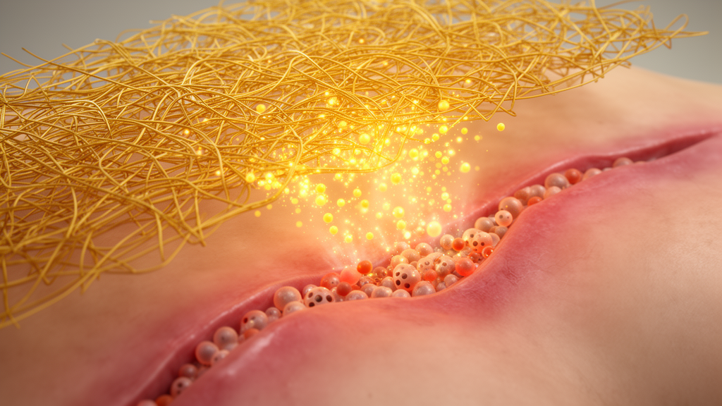
Chronic, infected wounds are among the most stubborn problems in modern medicine. Burns, traumatic injuries, and diabetic ulcers often stall in a hostile microenvironment defined by runaway inflammation, bacterial colonization, oxidative damage, and failed blood vessel growth. Now a team of researchers in China has built a single dressing that attacks all of these problems at once—and in animal tests it nearly closed infected wounds within two weeks. The material, described in Frontiers in Bioengineering and Biotechnology, weaves together polycaprolactone, carbon nanotubes, a mussel-inspired adhesive coating, and curcumin, the golden polyphenol extracted from turmeric, into one electroactive nanofibrous scaffold the authors call PCL/CNT-PDA@Cur.
The engineering starts with electrospinning, a technique that draws a polymer solution through an electrified needle to produce fibers hundreds of nanometers to a few micrometers across. The researchers dissolved polycaprolactone, an FDA-approved biodegradable polyester, in hexafluoroisopropanol, ultrasonically dispersed carbon nanotubes into the mix, and spun the blend into a randomly oriented fibrous mat. Scanning electron microscopy showed a uniform, bead-free network with high porosity and surface area—an architecture deliberately designed to mimic the native extracellular matrix that skin cells naturally inhabit. Because the fibers remain at the submicron scale even after modification, the scaffold keeps the flexibility and breathability a clinical dressing would need.
Carbon nanotubes give the mat its electrical personality. The team measured conductivity on the order of 10^-2 siemens per meter, a level that falls within the range of biological tissue conductivity. Crucially, the researchers emphasize that the scaffold is not a powered device: it is designed to conduct the endogenous electrical signals that wounds naturally generate, which are known to guide cell migration, proliferation, and differentiation. After the polydopamine coating was applied, conductivity was essentially unchanged, confirming that the modification did not sever the nanotube conductive pathways. The result is a passive scaffold that speaks the wound’s own bioelectric language.
That modification is where the mussel comes in. Polydopamine, formed by the self-polymerization of dopamine under weakly alkaline conditions, mimics the adhesive proteins that let mussels cling to wet rocks. When the team dipped the PCL/CNT mats into a dopamine solution for six hours, the coating transformed the surface chemistry: the static water contact angle plummeted from 135.46 degrees to 35.30 degrees, flipping the scaffold from strongly hydrophobic to markedly hydrophilic. Energy-dispersive X-ray spectroscopy detected nitrogen on the coated fibers, chemical fingerprints of the polydopamine layer. The catechol-rich coating then served as a molecular bridge for curcumin, binding the drug through pi-pi stacking and hydrogen bonding.
The binding chemistry solved one of curcumin’s oldest problems. The compound is pharmacologically attractive—anti-inflammatory, antioxidant, and antibacterial—but notoriously insoluble and rapidly metabolized. On unmodified scaffolds, curcumin loading efficiency was a modest 27.84 percent, and more than 60 percent of the loaded drug burst out within 24 hours. With the polydopamine layer, loading efficiency jumped to 71.38 percent, and release became sustained: roughly 25 percent in the first day, about 40 percent by 72 hours, then a slow trickle thereafter. That timing matters, because the inflammatory phase of wound healing typically spans 48 to 72 hours, precisely the window in which the scaffold keeps delivering its anti-inflammatory payload. The authors candidly note the trade-off: tight binding also limits total drug release, so future versions may need higher initial loading or degradable crosslinkers to balance burst suppression against release efficiency.
In laboratory cultures, the scaffold acted like a multi-tool for repair cells. L929 fibroblasts grown with scaffold extracts proliferated faster, spread into more extended morphologies, migrated more readily, and upregulated collagen types I and III, with the collagen ratio shifting toward the more mature profile seen in quality repair. Human umbilical vein endothelial cells showed elevated CD31 expression, upregulated VEGF, and formed denser, better-connected tubular networks on Matrigel—hallmarks of activated angiogenesis. Under hydrogen peroxide-induced oxidative stress, the scaffold protected fibroblasts, endothelial cells, and macrophages alike, cutting intracellular reactive oxygen species measured by DCFH-DA fluorescence and preventing the nuclear condensation and fragmentation of apoptosis seen in unprotected cells.
The immunomodulatory results may be the most consequential. Macrophages are the conductors of the wound orchestra, and their polarization state determines whether inflammation persists or resolves. When the researchers stimulated RAW264.7 macrophages with lipopolysaccharide and interferon-gamma, scaffolds carrying curcumin and polydopamine drove the cells away from the pro-inflammatory M1 phenotype—iNOS and TNF-alpha fell—toward the reparative M2 phenotype, with Arg-1 rising sharply. That M1-to-M2 switch is the critical transition between the inflammatory and proliferative phases of healing, and the dual action of polydopamine and curcumin appears to facilitate it directly.
Against bacteria, the scaffold killed more than 90 percent of both Staphylococcus aureus and Escherichia coli in plate-counting assays. Scanning electron microscopy revealed the mechanism in grim detail: bacterial cells on the curcumin-loaded scaffold shrank, ruptured, and leaked their contents, while control bacteria stayed smooth and intact. An ONPG permeability assay confirmed that bacterial membranes became leaky, and crystal violet staining showed the scaffold significantly suppressed S. aureus biofilm formation, a key driver of chronic infection and drug resistance. More intriguingly, quantitative PCR revealed that the scaffold downregulated RecA and LexA, the regulatory genes of the bacterial SOS response—the DNA damage repair program bacteria use to survive stress. The authors propose a synergistic model: membrane disruption kills directly, while SOS suppression strips bacteria of their repair machinery. SulA, an SOS effector gene, trended upward, which the team interprets as possible compensatory feedback. Whether curcumin directly inhibits RecA function, as prior work by Bellio and colleagues suggests, or whether scaffold-induced oxidative stress indirectly perturbs the pathway remains unresolved and is flagged for future study.
The decisive test came in rats. Forty Sprague-Dawley rats each received a 10-millimeter full-thickness skin defect contaminated with infection, covered with one of the scaffold variants or left untreated. By day 7, wounds under PCL/CNT-PDA@Cur were visibly smaller than all controls; by day 14 they were almost completely closed, with closure rates significantly exceeding the other groups at every measured time point. Bacterial cultures from wound tissue confirmed infection control in vivo. Immunofluorescence of day-7 tissue showed the same immune rewiring seen in vitro: CD206-positive M2 macrophages up, CD86-positive M1 macrophages down, TNF-alpha reduced, and IL-10 elevated. Histology told the rest of the story—thicker granulation tissue, the most abundant and orderly collagen deposition by Masson’s trichrome, elevated CD31-positive vascular area at day 7, and, uniquely among the groups, hair follicle-like structures in the regenerated skin, a marker of high-quality regeneration rather than mere scar closure.
The authors are appropriately careful about limits. Rat skin heals largely through contraction of the panniculus carnosus muscle, a mechanism that differs from human re-epithelialization, so the team cautions against direct clinical extrapolation and calls for validation in large animal models such as pigs, along with quantitative assessment of re-epithelialization and collagen maturity. Still, the study assembles a coherent therapeutic cascade—immune remodeling, then angiogenesis, then tissue regeneration—within one passive dressing that requires no external power, no antibiotics, and no growth factors. If the platform survives larger animal testing, turmeric chemistry, mussel adhesion, and carbon nanotube electronics may together offer a genuinely integrated answer to the infected wounds that medicine has struggled to close.
Subject of Research: A multifunctional curcumin-loaded electroactive nanofibrous scaffold for infected wound healing
Article Title: Polydopamine-mediated curcumin-functionalized electroactive nanofibrous scaffold accelerates infected wound healing via multifactorial microenvironment remodeling
Article References: Polydopamine-mediated curcumin-functionalized electroactive nanofibrous scaffold accelerates infected wound healing via multifactorial microenvironment remodeling. (n.d.). https://doi.org/10.3389/fbioe.2026.1959216
Image Credits: AI Generated
DOI: 10.3389/fbioe.2026.1959216
Keywords: curcumin, polydopamine, electrospinning, nanofibrous scaffold, wound healing, carbon nanotubes, bacterial SOS response, macrophage polarization, angiogenesis, oxidative stress, antibacterial biomaterials, skin regeneration
Cite Scienmag News
Denise Maddox. (October 16, 2026). Turmeric-Powered Nanofiber Scaffold Rewires Infected Wounds to Heal Faster. Scienmag. https://scienmag.com/turmeric-powered-nanofiber-scaffold-rewires-infected-wounds-to-heal-faster/
Denise Maddox. "Turmeric-Powered Nanofiber Scaffold Rewires Infected Wounds to Heal Faster." Scienmag, 16 October 2026, https://scienmag.com/turmeric-powered-nanofiber-scaffold-rewires-infected-wounds-to-heal-faster/. Accessed 16 October 2026.
Denise Maddox. "Turmeric-Powered Nanofiber Scaffold Rewires Infected Wounds to Heal Faster." Scienmag. October 16, 2026. https://scienmag.com/turmeric-powered-nanofiber-scaffold-rewires-infected-wounds-to-heal-faster/


