


Tetraspanins are four-transmembrane proteins best known for organizing cell membranes into specialized microdomains that recruit lipids and partner proteins. Over the past few years, several tetraspanins have also been shown to form ordered polymer assemblies, raising the possibility that these structures may act as functional scaffolds. Yet for TSPAN7, linked to intellectual disability, viral infection, diabetes, and cancer progression, the physical mechanism behind how it shapes and stabilizes tubular membrane structures remained unclear.
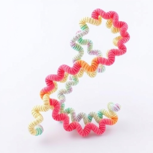
In a new study published in Vita, researchers show that TSPAN7 directly senses membrane curvature and polymerizes into a helical assembly on tubular membranes. Using a combination of live-cell imaging, in vitro reconstitution, and cryo-electron tomography, they uncover how individual TSPAN7 protomers self-assemble into a multi-start helix through two conserved interaction interfaces.
The team first observed that TSPAN7 accumulates on retraction fibers and tunneling nanotubes, and that elevating TSPAN7 expression enhances the formation of these structures. To test whether curvature recognition is intrinsic to TSPAN7, they reconstituted membrane tubes using giant unilamellar vesicles (GUVs). Without TSPAN7, the tubular network rapidly collapses into vesicles. With recombinant TSPAN7 present, TSPAN7 rapidly concentrates at the tubular network and preserves its curvature-dependent morphology.
Functional dynamics reinforced this model. Fluorescence recovery after photobleaching (FRAP) experiments showed TSPAN7 is largely immobile on retraction fibers and tunneling nanotubes, unlike other tetraspanin family members that display typical lateral diffusion. This immobilization persisted even after actin disruption with latrunculin A, suggesting the behavior arises from ordered polymerization rather than cytoskeletal coupling.
To visualize the molecular architecture, the authors used in situ cryo-EM and cryo-ET. TSPAN7 formed a highly ordered, right-handed helical structure on membrane tubes, with multiple protofilaments wrapping around the lipid membrane. Importantly, they identified two key protomer–protomer interfaces that drive assembly.
A double mutant designed to disrupt both interfaces failed to form spirals, regained mobility, and could not stabilize tubular membranes in vitro. Under microfluidic shear, cells expressing this spiral-deficient mutant exhibited exaggerated, deformation-prone membrane protrusions, whereas cells expressing wild-type TSPAN7 resisted tube constriction.
Together, the work supports a model in which TSPAN7 polymerizes into a membrane-embedded “transmembrane skeleton” that mechanically resists deformation. Notably, TSPAN7 and F-actin appear to occupy largely non-overlapping regions on protrusions, implying coordinated but distinct contributions to maintaining delicate membrane integrity.
By reframing a tetraspanin as an autonomous curvature sensor and intrinsic structural reinforcement, the study introduces a new conceptual class of membrane skeletal architecture. For viral and disease-relevant contexts where tubular conduits may facilitate spread and signaling, TSPAN7’s helical scaffold suggests a mechanistic lever that cells—and pathogens—may exploit.
Cite Scienmag News
Drew Townsend. (July 16, 2026). TSPAN7 Builds Transmembrane Skeleton, Stabilizing Tubular Membranes via Spiral Assembly. Scienmag. https://scienmag.com/tspan7-builds-transmembrane-skeleton-stabilizing-tubular-membranes-via-spiral-assembly/
Drew Townsend. "TSPAN7 Builds Transmembrane Skeleton, Stabilizing Tubular Membranes via Spiral Assembly." Scienmag, 16 July 2026, https://scienmag.com/tspan7-builds-transmembrane-skeleton-stabilizing-tubular-membranes-via-spiral-assembly/. Accessed 4 September 2026.
Drew Townsend. "TSPAN7 Builds Transmembrane Skeleton, Stabilizing Tubular Membranes via Spiral Assembly." Scienmag. July 16, 2026. https://scienmag.com/tspan7-builds-transmembrane-skeleton-stabilizing-tubular-membranes-via-spiral-assembly/


