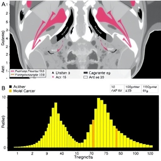
In an era dominated by the pursuit of precision medicine, the convergence of artificial intelligence and medical imaging stands at the forefront of transformative healthcare advances. A groundbreaking study published in BMC Cancer details a novel approach employing transformer models infused with habitat analysis from pretreatment ^18F-FDG PET imaging to predict overall survival outcomes in cervical cancer patients. This innovative methodology offers a promising horizon where personalized treatment strategies can be meticulously tailored, potentially revolutionizing prognostic accuracy in oncology.
Cervical cancer remains a significant global health challenge, with survival outcomes varying widely due to tumor heterogeneity and diverse biological behaviors. Traditional prognostic tools often fall short of capturing the nuanced microenvironment surrounding tumors. To address this, researchers from two medical institutions undertook a retrospective investigation involving 107 cervical cancer patients, applying advanced radiomic analyses to decode complex tumor habitats captured through ^18F-fluorodeoxyglucose positron emission tomography (PET).
Central to this study is the concept of “habitats” within and around tumors, which represent distinct radiological subregions characterized by unique metabolic and structural features. Utilizing a k-means unsupervised clustering algorithm, the researchers segmented the primary tumor and its immediate 4 mm peripheral peritumoral zone into four discrete habitats. This approach advances beyond conventional intratumoral focus by encompassing the peritumoral microenvironment, which plays a crucial role in tumor progression, metastasis, and therapeutic response.
Building upon these habitat delineations, a suite of transformer models was constructed to exploit radiomic features extracted from intratumoral, peritumoral, and habitat-specific subregions. Transformer architectures, originally conceived for natural language processing, have recently demonstrated profound capabilities in modeling complex relationships within diverse datasets. Their application here enables the exploration of spatial and metabolic patterns across different tumor habitats with heightened sensitivity and specificity.
Performance metrics reveal remarkable findings. Among the habitat-specific transformer models, the one analyzing habitat subregion 1 emerged as the most predictive, underscoring the critical biological relevance encoded within these microenvironments. When comparing individual models, the habitat-based transformer achieved an external validation AUC of 0.778, significantly surpassing models limited to intratumoral (AUC 0.714) or peritumoral (AUC 0.707) data alone. This differentiation confirms that capturing habitat heterogeneity lends superior prognostic granularity.
The study culminated in the development of an integrative transformer model combining intratumoral, peritumoral, and habitat features. This holistic framework attained an impressive validation AUC of 0.823, demonstrating not only enhanced predictive power but also robust calibration and clinical applicability. Such integrative modeling highlights the importance of multidimensional data fusion to fully unravel tumor behavior and patient survival probability.
Beyond pure statistical performance, decision curve analyses affirm the combined model’s potential to guide clinical decision-making. By effectively stratifying patients based on survival risk, this approach offers oncologists a powerful tool to identify individuals who might benefit from intensified therapeutic interventions or alternative treatment regimens. This advancement paves the way for precision oncology, where interventions are customized according to intricate tumor phenotypes rather than blunt clinical parameters.
The sophisticated methodology employed includes the extraction of high-dimensional radiomic features, capturing texture, intensity, and morphological characteristics of both tumor and surrounding tissue. When integrated within transformer networks, these features are contextualized in a spatially aware manner, enabling the models to detect subtle interactions and patterns indicative of aggressive tumor biology or favorable prognosis.
Importantly, this two-center retrospective study provides a broader validation framework, suggesting that the habitat-based transformer models possess generalizability across patient populations and imaging protocols. Such external validation is critical to assess the robustness and translational potential of AI-enabled prognostic tools before clinical adoption.
From a technological perspective, the choice of transformer architecture represents a significant leap in medical image analysis. Unlike traditional convolutional networks that focus locally, transformers employ self-attention mechanisms to weigh the relevance of distant features, capturing global contextual information. This fittingly resonates with the concept of tumor habitats, which may influence and be influenced by wider microenvironmental dynamics.
Furthermore, the study’s approach underscores the growing trend of integrating unsupervised machine learning techniques, like k-means clustering, to stratify biological heterogeneity without prior biases. Such unsupervised partitioning allows models to detect novel compartmentalization within tumor regions that might correspond to hypoxia, necrosis, or proliferative zones, expanding our biochemical and spatial understanding of cancer physiology.
Clinical implications stemming from these discoveries are profound. The ability to non-invasively prognosticate cervical cancer survival using advanced PET imaging combined with AI-driven habitat analysis could streamline patient management, reduce unnecessary toxic therapies, and focus resources on high-risk cases. Integrating this into routine workflows would mark a substantial leap toward personalized oncologic care.
In addition to its prognostic capacity, this study lays the groundwork for future research exploring dynamic changes within tumor habitats during and after treatment. Longitudinal monitoring with habitat-based transformers could reveal resistance mechanisms, therapeutic efficacy, or early recurrence, guiding adaptive clinical pathways in real time.
While the retrospective nature of the study and sample size provide initial encouraging evidence, prospective multicenter trials with larger cohorts are warranted to validate these findings. Optimizing habitat segmentation parameters and refining transformer architectures tailored to medical imaging modalities may further enhance prediction accuracy and clinical utility.
In summary, the convergence of habitat characterization in ^18F-FDG PET imaging and transformative AI architectures heralds a paradigm shift in cervical cancer prognosis. This innovative union empowers clinicians with unprecedented insight into tumor biology and survival outcomes, fostering strategic, patient-centric treatment plans. As artificial intelligence continues to permeate oncology, such integrative models stand as beacons of precision, promising improved survival and quality of life for patients worldwide.
Subject of Research:
Prediction of overall survival in cervical cancer patients using habitat-based transformer models applied to pretreatment ^18F-FDG PET imaging data.
Article Title:
Habitat-based transformer model in pretreatment ^18F-FDG PET imaging for predicting prognosis in cervical cancer: a two-center retrospective study.
Article References:
Lai, R., Tan, Q., Ding, C. et al. Habitat-based transformer model in pretreatment ^18F-FDG PET imaging for predicting prognosis in cervical cancer: a two-center retrospective study. BMC Cancer 25, 1515 (2025). https://doi.org/10.1186/s12885-025-14977-1
Image Credits:
Scienmag.com


