Surgeons reopening blocked arteries in the leg may soon be able to watch, in real time, how blood flow returns to the skin of the foot. A team at Sheffield Hallam University and Sheffield Teaching Hospitals NHS Foundation Trust has tested a thermal perfusion imaging system that maps microscopic circulation changes across the sole during endovascular procedures, reporting the results in Frontiers in Medical Technology.
The study was a prospective, exploratory MedTech development study designed to test the feasibility of the system in patients with chronic limb-threatening ischaemia. Of 23 participants recruited, video thermography was obtained in 21 patients admitted with moderate-severe disease complexity. Thirteen had disease confined to the femoral-popliteal segment, five had both femoral-popliteal and infrapopliteal disease, and three had infrapopliteal disease only.
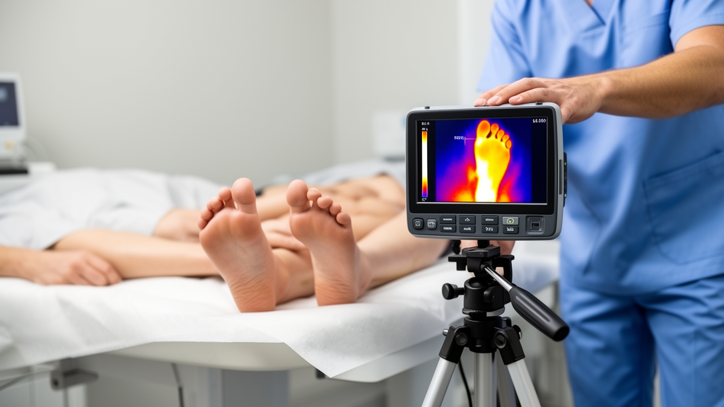
Intraprocedural video thermography was performed using a thermal camera with a resolution of 320 by 240 pixels. The system divided the plantar surface into five angiosomes, with regions of interest positioned within each. Initially, regions of interest were repositioned manually within each angiosome.
Subsequently, the analysis was automated using deep learning segmentation. Deep learning segmentation allowed the regions of interest to be tracked from the initial frame to the end of the procedure. Isotherms, described as thermal contours, were created and applied to the segmented surface. Changes in the percentage distribution of the isotherms overlying the treated and contralateral foot angiosomes provided quantitative, real-time measures during the procedure.
The results fell into two broad groups. Twelve patients were classified as plantar foot thermal perfusion responders and nine as non-responders. In responders, revascularisation produced a series of dynamic phases in which temperature rose in one or more angiosomes, and the hottest isotherms expanded significantly in response to the endovascular procedures, a difference the authors report at p = .002. In non-responders, no significant change in isotherm percentage distribution was observed during the intervention.
According to the research team, the system gives clinicians insight into microcirculatory changes at foot angiosomes in response to treatment through three parameters: temporal temperature profiles of each angiosome, real-time changes in the percentage distribution of isotherms across the plantar surface during the intervention, and angiosome-specific cutaneous perfusion responses to balloon inflation or stent deployment.
The authors suggest that locating the site of improved skin reperfusion has potential to predict treatment success for healing outcomes at the ulcer site, which they refer to as the woundosome.
Several caveats are worth keeping in view. This was an exploratory feasibility study in a modest sample of 21 patients with usable imaging, not a trial of clinical outcomes, and the conclusion the authors draw is that thermal perfusion imaging is feasible using the novel deep learning system. The suggestion that the technique could predict wound healing is framed as potential, not as a demonstrated result, and the study did not report follow-up healing data. The system was tested in a single centre’s patient series with moderate to severe disease complexity, so performance in other settings remains to be established.
Subject of Research: Feasibility of deep learning-assisted thermal perfusion mapping of foot angiosomes during endovascular revascularisation for chronic limb-threatening ischaemia
Article Title: Machine leaning assisted thermal reperfusion mapping reveals real-time changes in foot microcirculation during endovascular revascularisation
Article References: Childs, C., Doyle, K. J., Shenfield, A., & Goode, S. (2026). Machine leaning assisted thermal reperfusion mapping reveals real-time changes in foot microcirculation during endovascular revascularisation. Frontiers in Medical Technology, 8, Article 1950973. https://doi.org/10.3389/fmedt.2026.1950973
Image Credits: AI Generated
DOI: 10.3389/fmedt.2026.1950973
Keywords: thermal imaging, thermography, angiosomes, microcirculation, chronic limb-threatening ischaemia, endovascular revascularisation, deep learning, perfusion mapping, isotherms, peripheral artery disease, foot ulcers, medical technology
Cite Scienmag News
Ophelia Keating. (October 28, 2026). Thermal Camera Tracks Real-Time Foot Microcirculation During Leg Revascularisation. Scienmag. https://scienmag.com/thermal-camera-tracks-real-time-foot-microcirculation-during-leg-revascularisation/
Ophelia Keating. "Thermal Camera Tracks Real-Time Foot Microcirculation During Leg Revascularisation." Scienmag, 28 October 2026, https://scienmag.com/thermal-camera-tracks-real-time-foot-microcirculation-during-leg-revascularisation/. Accessed 29 October 2026.
Ophelia Keating. "Thermal Camera Tracks Real-Time Foot Microcirculation During Leg Revascularisation." Scienmag. October 28, 2026. https://scienmag.com/thermal-camera-tracks-real-time-foot-microcirculation-during-leg-revascularisation/