


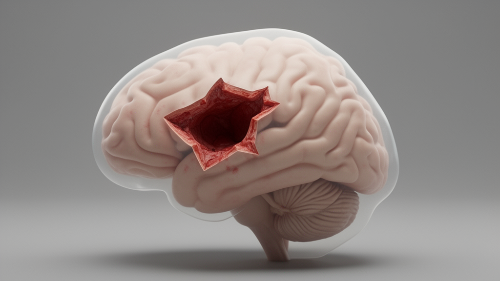
Every year, thousands of patients undergo surgery to remove gliomas—tumors that arise from the brain’s supporting cells—many of them located in so-called eloquent areas, regions that control speech, movement, or other critical functions. Surgeons operating in these zones walk a tightrope: remove as much tumor as possible while preserving the neural circuits that make a person who they are. Yet even with the most advanced imaging and intraoperative monitoring, a troubling question has persisted for decades. When a patient wakes up with a new neurological deficit, is it because a tiny stroke occurred during the operation, or is something else at play?
A new retrospective cohort study from a team of Italian neurosurgeons, neurologists, and neuroradiologists led by Francesco Guerrini of Fondazione IRCCS Policlinico San Matteo in Pavia offers a fresh clue. Published in the Journal of Neuro-Oncology, the research examines whether the shape of a lesion visible on postoperative diffusion-weighted imaging—DWI for short—can tell clinicians which patients are most likely to experience neurological worsening after eloquent-area glioma surgery. The answer, the researchers suggest, is more nuanced than a simple yes or no.
DWI is a magnetic resonance imaging technique that detects regions where water molecules cannot move freely, a hallmark of acute ischemic injury. When brain tissue is deprived of blood flow, cells swell and restrict the diffusion of water, producing a bright signal on DWI scans. For years, neurosurgeons have used postoperative DWI to identify silent strokes or small infarcts that might explain why a patient’s speech slurred or a limb weakened after tumor removal. But the technique has limitations. Bright spots on DWI do not always correspond to clinical deficits, and many patients who worsen neurologically show no obvious ischemic lesion at all.
The Italian team hypothesized that the morphology of these postoperative DWI lesions might matter. They classified lesions into two categories: sector-shaped, which appear as wedge-like areas of restricted diffusion, and rim-shaped, which form thin borders around the surgical cavity. Sector-shaped lesions, the researchers reasoned, might represent true arterial infarcts—blocks of tissue deprived of blood supply—while rim-shaped lesions could reflect a different phenomenon, perhaps related to surgical manipulation of the cortex surrounding the resection cavity rather than a genuine vascular occlusion.
To test this, the investigators analyzed 139 consecutive patients who underwent resection of supratentorial gliomas in eloquent areas, all classified as World Health Organization grade 2 through 4. Each patient received a postoperative MRI within 48 hours of surgery, allowing the team to categorize any DWI lesion by its shape. Neurological status was assessed at discharge and again at a follow-up visit four to six months later, providing both an early and a delayed picture of functional outcome.
The results were striking. DWI lesions appeared in 44 of the 139 patients, or 31.7 percent of the cohort. Of these, 31 were sector-shaped and 13 were rim-shaped. Overall, 59 patients—42.4 percent of the entire group—experienced neurological worsening at discharge. When the researchers looked at DWI positivity alone, without considering lesion shape, they found no significant association with worsening. But when they broke the lesions down by morphology, a clear pattern emerged. Sector-shaped DWI lesions were strongly associated with neurological decline, carrying a positive predictive value of 58.1 percent. In other words, if a patient had a wedge-shaped lesion on their postoperative scan, there was a better-than-even chance they would worsen neurologically. After adjusting for other variables in a multivariable model, sector-shaped lesions remained independently associated with worsening, with an odds ratio of 3.83. The confidence interval, however, was wide—ranging from 1.30 to 11.29—reflecting the modest sample size and suggesting that while the signal is real, its precise magnitude remains uncertain.
Rim-shaped lesions told a different story. These thin borders of restricted diffusion carried a positive predictive value of only 38.5 percent, which was essentially indistinguishable from the 37.9 percent risk of worsening observed in patients whose postoperative DWI was entirely negative. In a direct comparison between the two morphologies, the difference did not reach statistical significance, though the trend favored the idea that sector-shaped lesions are the more clinically meaningful finding. This distinction matters because it suggests that not all bright spots on a postoperative DWI scan are created equal. A rim of restricted diffusion hugging the cavity wall may be a benign epiphenomenon of surgery, while a wedge-shaped lesion in the territory of a perforating artery may signal a true ischemic event with real functional consequences.
Perhaps the most sobering finding of the study is what it reveals about the limits of DWI itself. Sixty-one percent of all neurological worsening in this cohort occurred in patients whose postoperative scans showed no DWI lesion whatsoever. This means that the majority of early postoperative decline after eloquent-area glioma surgery cannot be explained by ischemia visible on DWI. The researchers suggest that non-ischemic mechanisms—such as mechanical manipulation of subcortical pathways, edema, vascular dysregulation, or transient metabolic disruption—may be responsible. This finding aligns with a growing body of literature suggesting that surgically acquired deficits often arise from disruption of white matter tracts rather than from overt stroke, and it underscores the need for imaging modalities or biomarkers that can detect these subtler forms of injury.
The study also identified two additional factors associated with neurological outcome. Awake craniotomy, a technique in which the patient is woken during surgery to perform language or motor tasks, was independently associated with worsening, a finding that may reflect the inherent difficulty of operating in eloquent cortex rather than a flaw in the technique itself. Meanwhile, tumors located in the frontal lobe carried a lower risk of worsening compared with those in other eloquent regions, possibly because frontal language and motor networks possess greater redundancy or because surgical corridors in the frontal lobe are more forgiving. At the four-to-six-month follow-up, only nine patients—6.5 percent of the cohort—still had a deficit, and all of them had IDH-wildtype glioblastoma, the most aggressive and least metabolically favorable glioma subtype. Persistent deficit was also associated with a decrease of at least 50 percent in intraoperative evoked potentials, a neurophysiological monitoring signal that reflects the integrity of motor or sensory pathways during surgery.
These findings carry important implications for the field of neuro-oncology. First, they suggest that radiologists and surgeons should pay close attention to lesion morphology when interpreting postoperative DWI in glioma patients, rather than simply reporting the presence or absence of restricted diffusion. A sector-shaped lesion warrants closer clinical scrutiny, while a rim-shaped lesion may be less alarming than its appearance suggests. Second, the high proportion of worsening without DWI ischemia reinforces the idea that function-guided surgery—whether performed awake or asleep—exerts its effects on the brain through mechanisms that standard MRI cannot fully capture. Third, the association between persistent deficit and IDH-wildtype glioblastoma highlights the biological differences between glioma subtypes and suggests that patients with these aggressive tumors may have less capacity for functional recovery after surgery. Finally, the authors caution that their findings require confirmation in larger, prospective series, given the retrospective design, the modest number of patients with each lesion morphology, and the wide confidence intervals around some of their estimates. Still, the study represents a meaningful step toward a more refined understanding of what happens to the brain during eloquent-area glioma surgery, and it offers clinicians a new lens—quite literally—through which to interpret the postoperative scan.
Subject of Research: Postoperative diffusion-weighted imaging lesion morphology and neurological outcome after eloquent-area glioma surgery
Article Title: Postoperative DWI morphology and neurological worsening after eloquent-area glioma surgery: a retrospective cohort study
Article References: Guerrini, F., D’Agata, F., Musella, V., Vitali, A., Giuri, A., Caporrimo, M. C., Chiapparini, L., & Spena, G. (2026). Postoperative DWI morphology and neurological worsening after eloquent-area glioma surgery: a retrospective cohort study. Journal of Neuro-Oncology, 180(2), Article 23. https://doi.org/10.1007/s11060-026-05837-4
Image Credits: AI Generated
DOI: 10.1007/s11060-026-05837-4
Keywords: glioma surgery, diffusion-weighted imaging, DWI lesion morphology, eloquent cortex, neurological worsening, awake craniotomy, intraoperative neurophysiology, postoperative MRI, IDH-wildtype glioblastoma, surgical outcomes, neuro-oncology, brain tumor
Cite Scienmag News
Nathaniel Bowman. (October 17, 2026). The Shape of a Postoperative Brain Lesion May Predict Neurological Decline After Glioma Surgery. Scienmag. https://scienmag.com/the-shape-of-a-postoperative-brain-lesion-may-predict-neurological-decline-after-glioma-surgery/
Nathaniel Bowman. "The Shape of a Postoperative Brain Lesion May Predict Neurological Decline After Glioma Surgery." Scienmag, 17 October 2026, https://scienmag.com/the-shape-of-a-postoperative-brain-lesion-may-predict-neurological-decline-after-glioma-surgery/. Accessed 17 October 2026.
Nathaniel Bowman. "The Shape of a Postoperative Brain Lesion May Predict Neurological Decline After Glioma Surgery." Scienmag. October 17, 2026. https://scienmag.com/the-shape-of-a-postoperative-brain-lesion-may-predict-neurological-decline-after-glioma-surgery/


