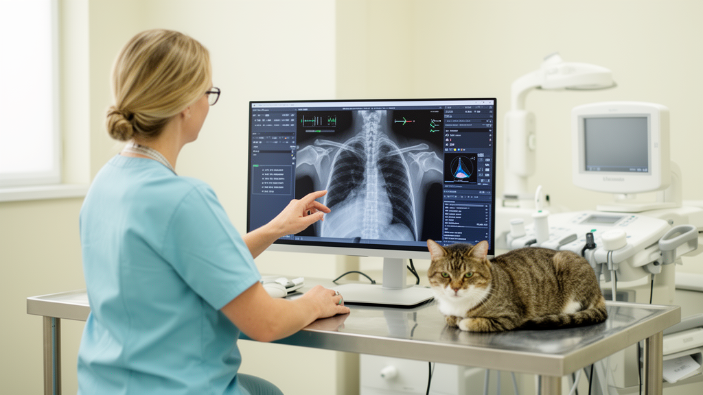
A systematic review of 57 studies involving dogs and cats found that convolutional neural networks and long short-term memory networks can produce high diagnostic performance in controlled research settings, but only six studies used independent external validation. The evidence was concentrated in oncology, cardiology and pulmonology, while limited independent testing and inconsistent reporting of patient-level data separation left the models’ reliability in routine veterinary practice uncertain.
The researchers searched Scopus and the Web of Science Core Collection on April 23, 2026, for peer-reviewed English-language, open-access journal articles published from 2021 through 2026. They included primary empirical studies of canine and feline diagnostic or decision-support systems based on CNNs or LSTMs while excluding work centered on survival prediction, treatment efficacy, traditional machine learning, qualitative studies, reviews, livestock, wildlife and single-animal case reports. The initial search identified 225 records. After 67 duplicates were removed, 93 records were excluded during title-and-abstract screening and one report could not be retrieved. Full-text assessment excluded seven more studies, leaving 57 studies for synthesis.
The review followed PRISMA 2020 guidelines and organized its eligibility criteria around a PICO-CS framework covering population, intervention, comparison, outcome, context and study design. Although the search strategy, criteria, research questions and analytical framework were established before the search and extraction, no prospective review protocol was registered in a public registry. Comparison with conventional procedures was addressed qualitatively through reference standards such as histopathological confirmation and specialist consensus rather than through a database search designed to compare neural networks directly with clinical methods. Screening and extraction were conducted independently by three authors, with disagreements resolved by discussion.
Eleven clinical domains were represented in the final corpus. Oncology was the largest, accounting for 28.1% of the studies, followed by cardiology at 15.8% and pulmonology at 12.3%. Investigated conditions included mammary tumors, mast cell tumors, lymphomas and osteosarcoma; cardiomegaly, myxomatous mitral valve disease and feline hypertrophic cardiomyopathy; pleural effusion, pulmonary abnormalities and coccidioidomycosis. Other applications involved cataracts and retinal detachment, hip and elbow dysplasia, blood-cell morphology, babesiosis, skin lesions, seizure prediction, activity and separation-anxiety monitoring, chronic kidney disease and estrus-cycle classification. Dogs appeared more frequently than cats, and feline studies were particularly concentrated in cardiology and pulmonology.
Architectural choices were closely associated with the clinical data modality. CNNs predominated in applications involving spatial information, including radiographs, clinical photographs, ultrasonography, CT, MRI and microscopic imagery. ResNet was the most frequently used family, appearing across six of the seven evaluated modalities, particularly radiographs and microscopic images. U-Net was used for tumor-region and cardiac-silhouette segmentation; YOLO and R-CNN frameworks supported object detection; Inception was applied to classification; and Vision Transformers were used in histopathology and clinical photography. Clinical-photography studies also made relatively extensive use of EfficientNet. LSTMs and recurrent neural networks were confined to sequential data in the reviewed literature, including epileptic seizure recordings, accelerometry from wearables, mitral murmurs and kinematic gait analysis. Some hybrid systems converted temporal signals into spectrograms for subsequent processing by two-dimensional CNNs.
Binary classification produced the strongest reported performance. Across the relevant studies, accuracy ranged from 0.82 to 0.99, area under the receiver operating characteristic curve from 0.69 to 0.99, F1-scores from 0.85 to 0.99 and sensitivity from 0.82 to 0.97. High values were reported in work on cardiomegaly detection from thoracic radiographs and tumor classification from microscopic images. Multi-class tasks showed accuracy from 0.78 to 0.99, F1-scores from 0.68 to 0.95 and macro-F1 scores from 0.68 to 0.94. The review associated this broader variation with differences in dataset characteristics, class distribution, task complexity and validation protocols, but the synthesized evidence did not establish any single factor as the definitive cause.
Other diagnostic tasks were evaluated with task-specific measures that could not be compared directly with classification accuracy. For object detection, mean average precision ranged from 0.87 to 0.97, F1-scores ranged from 0.75 to 0.84 and sensitivity ranged from 0.91 to 0.97. Applications included detecting mitotic figures in tissue images and pododermatitis in clinical photographs. For segmentation, Dice similarity coefficients ranged from 0.58 to 0.97, while intersection-over-union values ranged from 0.58 to 0.95. Tumor-region segmentation in histopathology and cardiac-silhouette delineation on radiographs produced some of the lower values. Temporal studies were more heterogeneous: seizure prediction, behavioral monitoring and related tasks reported sensitivity values from 0.62 to 1.00. Regression studies included wound-volume quantification with mean absolute percentage error of 5% to 30% and cardiac-radiograph assessment with a mean bias of −0.09 vertebrae.
Validation practices limit how confidently those figures can be interpreted. Of the 57 studies, 54 used a standard hold-out split and 26 used k-fold cross-validation, but only six, or 10.5%, evaluated performance on external datasets from independent institutions. Among studies handling multiple images, frames or observations from each animal, only seven explicitly reported splitting data at the patient level. If observations from the same animal appear in both training and testing, they can make performance appear stronger than it would be on unseen patients. The review’s six-criterion assessment nevertheless assigned high composite methodological-quality scores to 55 studies and moderate scores to two. Model descriptions and task-specific performance measures were generally well documented, whereas independent validation and safeguards against subject-level data dependence were reported less often.
Primary veterinary care was the most common intended setting, with models presented as decision-support aids for clinicians rather than substitutes for professional evaluation. Proposed uses included screening thoracic radiographs for cardiac or pulmonary abnormalities, quantifying vertebral heart size and joint structures, classifying skin and retinal conditions, and supporting emergency triage. Specialized laboratories were the second major context, especially for histopathology, cytology and hematology, while radiology suites, emergency services and remote wearable monitoring appeared less often. The research was geographically concentrated in high-income countries, including the United States, South Korea and several European nations, with sparse participation from Turkey, India, Thailand, Brazil and Algeria. This distribution describes where studies were conducted rather than evidence of a geographic decline over time.
Computational requirements add another obstacle to point-of-care use. Some models were designed for mobile or edge devices, but others depended on dedicated graphics processors, cloud services or high-performance computing systems. Many studies did not specify minimum hardware requirements, inference times or execution costs, making it difficult to determine whether they could run on conventional equipment in resource-constrained clinics. The heterogeneity of populations, datasets, tasks, metrics and validation procedures also prevented meta-analytic pooling. The review therefore supports further evaluation rather than establishing that the surveyed systems are ready for routine deployment. The authors call for prospective multicenter studies, independent external validation, representative open-access datasets, transparent reporting of validation procedures, inference performance and hardware needs, and appropriate regulatory assessment before clinical use.
Subject of Research: Systematic review of CNN- and LSTM-based diagnostic decision support in dogs and cats
Article Title: Deep learning for diagnostic and clinical decision support in companion animals: a systematic review of CNN- and LSTM-based approaches
Article References: Guevara-Flores, J. J., López-Rojas, J., & Vásquez–Coronel, J. A. (2026). Deep learning for diagnostic and clinical decision support in companion animals: a systematic review of CNN- and LSTM-based approaches. Frontiers in Veterinary Science, 13, Article 1952589. https://doi.org/10.3389/fvets.2026.1952589
Image Credits: AI Generated
DOI: 10.3389/fvets.2026.1952589
Keywords: veterinary artificial intelligence, diagnostic imaging, convolutional neural networks, long short-term memory, companion animals, external validation, clinical deployment, canine medicine, feline medicine, Deep, learning, diagnostic
Cite Scienmag News
William Thompson. (October 23, 2026). Systematic review finds limited validation of AI diagnosis in dogs and cats. Scienmag. https://scienmag.com/systematic-review-finds-limited-validation-of-ai-diagnosis-in-dogs-and-cats/
William Thompson. "Systematic review finds limited validation of AI diagnosis in dogs and cats." Scienmag, 23 October 2026, https://scienmag.com/systematic-review-finds-limited-validation-of-ai-diagnosis-in-dogs-and-cats/. Accessed 23 October 2026.
William Thompson. "Systematic review finds limited validation of AI diagnosis in dogs and cats." Scienmag. October 23, 2026. https://scienmag.com/systematic-review-finds-limited-validation-of-ai-diagnosis-in-dogs-and-cats/