


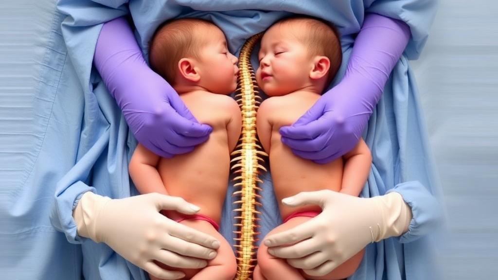
In a remarkable surgical achievement that is already drawing international attention, a team of doctors at a resource-limited tertiary care hospital has successfully separated a pair of pygopagus conjoined twin girls who shared not only fused soft tissue at the sacrum but also a common dural envelope and conjoined lower spinal cord elements. The case, documented in a recent clinical report, stands out because the operation was performed without the intraoperative neurophysiological monitoring technology that is considered the gold standard in well-resourced centers. Instead, the surgical team relied on meticulous microsurgical technique, fixed anatomical landmarks, and a disciplined, stepwise approach to nerve root identification. Both infants survived the procedure, recovered without major complications, and were discharged with preserved motor function, sensation, and sphincter control, offering a powerful demonstration that complex pediatric neurosurgery can succeed even where advanced equipment is scarce.
Conjoined twins arise from aberrant embryogenesis in a monozygotic, monochorionic pregnancy, in which the developing embryos fail to separate completely. They are extraordinarily rare, with an estimated incidence of one in 100,000 to 250,000 births, and roughly half of affected pregnancies end in stillbirth. The condition occurs more frequently in females, at a ratio of approximately three to one. Classification depends on the site of fusion: thoracopagus twins are joined at the chest, omphalopagus at the abdomen, ischiopagus at the pelvis, craniopagus at the head, and pygopagus twins at the sacrum or rump. Pygopagus twins account for only about 6 to 19 percent of all conjoined twin cases, making them one of the rarer varieties. In pygopagus fusion, the twins typically share portions of the gastrointestinal and genitourinary systems, and in a small minority of cases the neural elements are involved as well. Of the pygopagus separations reported in the medical literature, shared dura has been documented in 17 cases and shared spinal cord in only 10, underscoring just how unusual the anatomy in this new case truly was.
The twins in this report were discovered during a routine prenatal ultrasound at 20 weeks of gestation, allowing the mother and her obstetricians to plan carefully for the delivery. Her antenatal course remained otherwise uneventful, and the babies were born by preplanned lower segment cesarean section at a government medical hospital, with a combined birth weight of 3.6 kilograms. The infants were fused at the lower body in the sacral region by a soft tissue bridge measuring roughly 10 centimeters across the skin. They were referred to a tertiary medical college two days after birth, where they were designated Twin A and Twin B. Baseline investigations showed stable physiological parameters in both infants, and echocardiography confirmed normal cardiac anatomy and function in each, with no shunt anomalies and normal pulmonary artery pressures. Abdominal ultrasound detected no significant abnormalities, meaning the twins did not share vital visceral organs, a factor that substantially improved their surgical prognosis.
When a multidisciplinary board of pediatric surgeons, neurosurgeons, pediatricians, anesthesiologists, and a dedicated pediatric clinical nutritionist convened to evaluate the pair, they reached a critical conclusion: the infants were simply too small and too underweight to withstand the physiological trauma of a prolonged separation surgery. Moreover, the skin and subcutaneous tissue surrounding the conjoined pelvic junction were judged insufficient for tension-free primary closure. Rather than rushing to the operating room, the board opted for a delayed elective approach, instituting a structured nutritional rehabilitation program supervised by the clinical nutritionist. The twins received maternal breastfeeding supplemented with calorie-dense nutrition, with a target weight gain velocity of 20 to 30 grams per day per infant. Over the following months, their combined weight climbed to 8.9 kilograms, and biochemical markers confirmed that they had built sufficient physiological reserves to tolerate intraoperative blood loss, lengthy anesthesia, and the hypercatabolic stress of post-surgical healing.
The delay also served a second, equally important purpose: it allowed natural somatic growth to generate the redundant skin needed for reconstruction. The team had formally considered inserting mechanical tissue expanders, devices commonly used to stretch skin before complex closures. They ultimately rejected this option for three reasons: stringent institutional resource limitations, the heightened risks of expander erosion, hematoma, and catastrophic implant infection given the proximity to the diaper and perineal region, and the encouraging clinical evidence that spontaneous growth was producing sufficient cutaneous redundancy on its own. Surgeons serially assessed the pinch-laxity and dermal mobility of the lateral gluteal and flank skin relative to fixed skeletal landmarks, including the sacrococcygeal junction and the posterior superior iliac spines. Robust capillary refill times of under two seconds and the absence of scar tethering confirmed healthy microvascular perfusion, and by the time of surgery the natural tissue creep had generated enough lateral skin folds to permit primary local flap reconstruction without any synthetic prostheses.
Neuroimaging proved decisive in mapping the twins’ shared anatomy. A computed tomography scan with three-dimensional reconstruction at three months of age revealed that each twin had an independent, anatomically normal cervical, thoracic, and lumbar spine, with no osseous bridging and skeletal contact limited to the lower sacrococcygeal region. Magnetic resonance imaging of the pelvis then delivered the finding that defined the case: the sacrum was deficient in both infants, and the distal spinal cords and dural sacs converged and fused near the sacrococcygeal level within the soft tissue bridge. The thoracic and abdominal visceral compartments remained entirely separate. This combination of shared spinal cord and shared dura is exceptionally rare in pygopagus twins and dramatically raises the stakes of separation, since any misjudgment during cord division risks permanent neurological injury to one or both children. After reviewing the imaging, the surgical board scheduled the operation, though it was postponed once more when Twin A developed pneumonia, ultimately taking place six months after birth, which coincidentally aligned with the internationally recommended window of four to twelve months for planned elective separation.
The operation itself showcased how surgical precision can substitute for technology. Because advanced intraoperative neurophysiological monitoring, including somatosensory evoked potentials, motor evoked potentials, and electromyographic nerve root stimulation, was unavailable, neural preservation depended entirely on high-magnification operating microscopy and anatomical landmarks. The infants were positioned in a synchronized lateral decubitus position, and the team designed a modified curvilinear zigzag, interlocking S-pattern incision over the posterior bridge, a geometry chosen to distribute skin tension evenly, prevent straight-line scar contracture over the sacrum, and optimize flap interdigitation for each twin. Subcutaneous flaps were elevated down to the paraspinal fascia, and the sacrococcygeal junction served as the principal anchor for the exposure. Under high-power magnification, the fused thecal envelope was incised longitudinally along the midline raphe, opening the common dural chamber.
Inside the shared thecal sac, the surgeons traced each nerve root systematically, both proximally toward its origin on the cord and distally toward its corresponding neural foramen, confirming bilaterally and visually that every root belonged unequivocally to its respective infant before any division. With gentle blunt micro-dissection and continuous saline irrigation, the conjoined lower cord elements were separated without sacrificing functioning neural tissue from either twin, and the thecal sac was partitioned into two distinct envelopes. Each baby was then moved to a separate operating table, where the team repaired the dura under the microscope using 6-0 polypropylene sutures in a watertight fashion, reinforced the closure with a fascial layer, and confirmed the absence of cerebrospinal fluid leakage with a Valsalva maneuver before closing the wounds in layers. Cerebrospinal fluid leakage is a historically frequent and dangerous complication in pygopagus separations involving dural sharing, making this watertight closure one of the operation’s most consequential technical victories.
The postoperative course was largely uneventful. Apart from a bout of pneumonia in Twin B on the third day, which was treated successfully, neither infant developed surgical site infections, wound dehiscence, flap necrosis, or cerebrospinal fluid leaks. Detailed neurological examinations before discharge revealed active, symmetric, spontaneous movements of both lower extremities in each infant, normal muscle tone across the hip flexors, quadriceps, and gastrocnemius muscles, and brisk, bilaterally equal deep tendon reflexes. Sensory withdrawal responses were intact across all lumbar and sacral dermatomes from L1 to S3, and both twins displayed robust anal wink reflexes, confirming preservation of the S2 to S4 sacral autonomic and pudendal pathways that govern bowel and bladder control. Both infants demonstrated normal spontaneous fecal evacuation and independent voluntary voiding. They were released on the twelfth postoperative day with basic vitamin supplements and are now reportedly healthy and living independently.
The outcome carries weight well beyond this single hospital. Survival rates for conjoined twins after separation range from roughly 16.7 to 50 percent depending on the type of operation and whether it is elective or emergency, and mortality climbs sharply when neurological structures are shared, since it is exceptionally difficult for both twins to survive the division of a joined spinal cord. The authors of the report are candid about the limitations of their approach: nutritional and skin assessments relied on clinical expertise and biochemical parameters rather than validated scoring tools or perfusion devices, no real-time electrophysiological feedback was available during cord division, and follow-up assessments were clinical rather than urodynamic or electrodiagnostic. Even so, the case demonstrates that three surgical strategies, fixed skeletal anchoring, stepwise magnified micro-dissection, and individual rootlet verification before dural division, can substitute meaningfully for expensive neuromonitoring. For surgical teams across the developing world, where conjoined twin separations are often deemed impossible without technology-rich environments, this successful delayed elective separation offers both a practical roadmap and a compelling proof of concept.
Subject of Research: Delayed elective surgical separation of pygopagus conjoined twins with shared spinal cord and dura
Article Title: Successful Delayed Elective Separation of Pygopagus Conjoined Twins With Shared Spinal Cord and Dura in a Resource‐Limited Setting: A Case Report
Article References: Wahid, Z. U. M., Chowdhury, S. I., Islam, N., & Mifty, S. K. (2026). Successful Delayed Elective Separation of Pygopagus Conjoined Twins With Shared Spinal Cord and Dura in a Resource‐Limited Setting: A Case Report. Clinical Case Reports, 14(10), Article e73657. https://doi.org/10.1002/ccr3.73657
Image Credits: AI Generated
DOI: 10.1002/ccr3.73657
Keywords: conjoined twins, pygopagus, spinal cord, separation surgery, neurosurgery, pediatric surgery, microsurgery, resource-limited settings, dural repair, nutritional optimization, case report, tissue expansion
Cite Scienmag News
Ophelia Keating. (October 5, 2026). Surgeons Separate Conjoined Twins Sharing a Spinal Cord Without Advanced Equipment. Scienmag. https://scienmag.com/surgeons-separate-conjoined-twins-sharing-a-spinal-cord-without-advanced-equipment/
Ophelia Keating. "Surgeons Separate Conjoined Twins Sharing a Spinal Cord Without Advanced Equipment." Scienmag, 5 October 2026, https://scienmag.com/surgeons-separate-conjoined-twins-sharing-a-spinal-cord-without-advanced-equipment/. Accessed 5 October 2026.
Ophelia Keating. "Surgeons Separate Conjoined Twins Sharing a Spinal Cord Without Advanced Equipment." Scienmag. October 5, 2026. https://scienmag.com/surgeons-separate-conjoined-twins-sharing-a-spinal-cord-without-advanced-equipment/


