


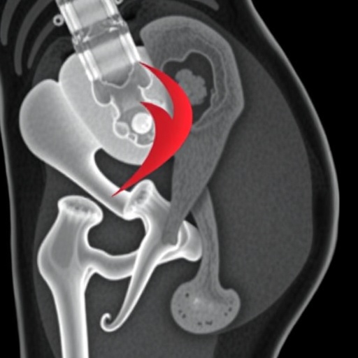
For reconstructive surgeons, one of the most important decisions often happens before an operation begins: identifying the tiny blood vessels that will keep a transplanted piece of tissue alive. A new study in Scientific Reports examines how different imaging technologies perform when mapping these vessels in the anterolateral thigh, a region widely used as a source of tissue for complex reconstructive surgery. By comparing imaging methods within the same individuals, the researchers sought to clarify how reliably each technique can reveal the anatomy of anterolateral thigh perforators and support safer, more predictable surgical planning.
The anterolateral thigh, commonly abbreviated ALT, is a remarkably versatile donor site. Surgeons can remove skin, fat, fascia and, when necessary, portions of muscle from the thigh and transfer them to another part of the body while preserving the leg’s overall function. These tissue transfers, known as free flaps, are used to repair injuries, remove the consequences of cancer surgery and reconstruct areas such as the head and neck, breast, hands and lower limbs. Their success depends on a vascular connection: arteries and veins must supply the tissue and drain it after transplantation. The small arteries that pass from the deeper muscles toward the skin are called perforators, and finding them accurately is central to the procedure.
Perforator anatomy is difficult to predict from one person to another. Some vessels travel directly through the muscle, while others follow an intermuscular or septocutaneous route between muscle groups. Their diameter, length, branching pattern and relationship to the surrounding tissue can vary substantially. A vessel that appears ideal on one scan may prove too short, too narrow or technically difficult to dissect once surgery begins. Preoperative mapping is therefore intended to reduce uncertainty, guide the incision and help surgeons select a perforator with a suitable course and caliber before the first cut is made.
The study by Andreas Geierlehner, Rainer E. Horch, Matthias Uder and colleagues is notable because it uses an intraindividual comparison. Rather than comparing separate groups of patients who underwent different scans, the investigators evaluated imaging modalities in the same subjects. This approach limits the influence of individual anatomical variation, which is especially important in perforator surgery. A comparison performed within each person can reveal whether one method consistently identifies the same vessels as another, whether the techniques provide different types of information and where disagreements may arise.
The imaging technologies involved in perforator mapping each rely on a different physical principle. Computed tomography angiography, or CTA, uses X-rays and an injected contrast agent to produce high-resolution images of blood vessels and the surrounding anatomy. It can display the three-dimensional course of a perforator from its source artery through muscle and toward the skin, making it useful for surgical road maps. Its disadvantages include exposure to ionizing radiation and the need for iodinated contrast, which can be unsuitable for some patients.
Magnetic resonance angiography, or MRA, creates vascular images using magnetic fields and radiofrequency signals. Depending on the protocol, it can provide excellent soft-tissue contrast and may avoid ionizing radiation, an advantage when repeated imaging is considered. However, MRA can be more sensitive to motion, may require longer acquisition times and does not always depict very small vessels in exactly the same way as CTA. In addition, the use of gadolinium-based contrast agents may be limited in patients with certain forms of kidney disease. These trade-offs make direct clinical comparison important rather than assuming that one technology is universally superior.
Ultrasound adds another dimension to the assessment. With color Doppler and power Doppler techniques, clinicians can observe blood moving through a vessel in real time. The examiner can trace a perforator, measure its flow and evaluate its position relative to the skin and muscle while applying the probe. Duplex ultrasound can also provide information about blood-flow direction and velocity. Yet the method is operator-dependent, and deep or unusually positioned vessels may be difficult to follow. Its usefulness can depend on the patient’s anatomy, the quality of the equipment and the experience of the person performing the examination.
For surgeons, the most valuable result is not necessarily a single “winning” scan, but a clear understanding of what each modality can contribute. A cross-sectional technique such as CTA or MRA can provide a broad anatomical map, showing the source vessels and their relationship to muscles, fascia and bone. Ultrasound can then help confirm the location of a superficial perforator and assess its flow immediately before surgery. When the findings agree, confidence in the operative plan increases. When they do not, the discrepancy may signal the need for additional imaging or a different flap design.
The research also speaks to a larger movement in reconstructive medicine: replacing approximate anatomical expectations with individualized vascular maps. Modern free-flap surgery increasingly depends on patient-specific planning, especially as surgeons attempt to preserve muscle, shorten operating time and tailor tissue transfers to complex defects. Accurate perforator mapping can reduce unnecessary dissection and may help prevent damage to vessels that are not selected for the flap. It can also support the design of thinner or more precisely shaped tissue transfers, which may improve both functional recovery and cosmetic results.
The findings are relevant beyond one donor site. Similar imaging questions arise whenever surgeons rely on small perforating vessels, including deep inferior epigastric perforator flaps used in breast reconstruction, profunda artery perforator flaps from the thigh and other muscle-sparing procedures. The study’s intraindividual design offers a useful framework for evaluating imaging pathways in these settings. Rather than treating imaging as a routine preliminary step, it positions vascular mapping as a measurable component of surgical decision-making—one that can be optimized according to the patient, the anatomy and the clinical goal.
At a time when medical imaging is often promoted through claims of greater speed, resolution or artificial intelligence, the study provides a more grounded message: the best technology is the one that answers the surgeon’s specific anatomical question with the least unnecessary burden for the patient. CTA, MRA and Doppler ultrasound each reveal different aspects of perforator anatomy, and their value may depend on whether the priority is three-dimensional vessel tracking, soft-tissue characterization, radiation avoidance or real-time flow assessment. By comparing modalities within the same individuals, Geierlehner and colleagues bring the discussion closer to everyday surgical reality. Their work reinforces a principle that is increasingly shaping personalized reconstruction: better maps can lead to better operations, but only when the map accurately reflects the unique vascular landscape beneath each patient’s skin.
Cite Scienmag News
Courtney Benton. (August 24, 2026). Study Compares Imaging Methods for Mapping Anterolateral Thigh Perforators. Scienmag. https://scienmag.com/study-compares-imaging-methods-for-mapping-anterolateral-thigh-perforators/
Courtney Benton. "Study Compares Imaging Methods for Mapping Anterolateral Thigh Perforators." Scienmag, 24 August 2026, https://scienmag.com/study-compares-imaging-methods-for-mapping-anterolateral-thigh-perforators/. Accessed 4 September 2026.
Courtney Benton. "Study Compares Imaging Methods for Mapping Anterolateral Thigh Perforators." Scienmag. August 24, 2026. https://scienmag.com/study-compares-imaging-methods-for-mapping-anterolateral-thigh-perforators/


