


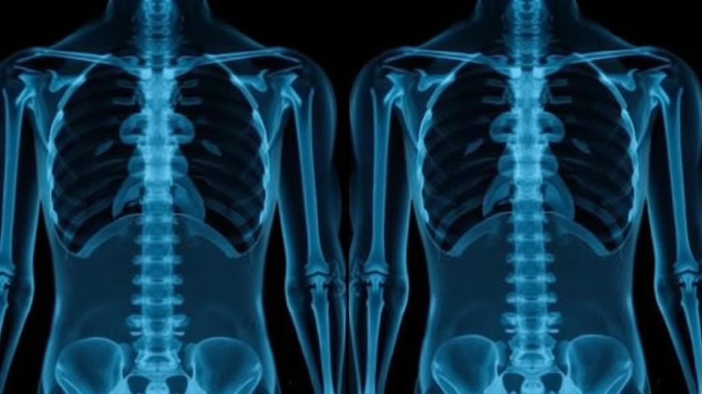
For millions of people worldwide, low back pain that shoots down the leg is the unmistakable signature of a herniated disc in the lumbar spine. Most of these cases settle with rest, physical therapy, and time. But a subset of patients harbor a herniation so large that it crushes more than half of the spinal canal, dramatically raising the odds that conservative treatment will fail and surgery will become necessary. The problem, according to a new study from Thailand, is that the only reliable way to measure that compression—magnetic resonance imaging—is exactly the resource that many clinics around the world simply do not have. Now researchers report that a simple measurement taken from an ordinary standing X-ray may help identify which patients deserve a fast track to the MRI scanner.
The study, published in Frontiers in Musculoskeletal Disorders by Suttinont Surapuchong of Lerdsin General Hospital and Krit Pongpirul of Chulalongkorn University, examined whether parameters routinely measured on standing whole-spine radiographs are associated with the severity of spinal canal compromise as defined by MRI. The question is deceptively practical. MRI is the undisputed gold standard for visualizing displaced disc material and quantifying how much it compresses the canal, but it is costly, often scarce in primary and secondary care settings, and typically withheld for four weeks or more while conservative treatment is tried first. Standing X-rays, by contrast, are cheap, universally available, and usually the very first imaging test a back-pain patient receives. If certain X-ray measurements could hint at severe compression, clinicians in resource-limited environments might triage referrals more intelligently.
To test the idea, the team conducted a retrospective cross-sectional study at Lerdsin Hospital in Bangkok, drawing on patients seen between June 2014 and January 2024. They identified candidates using the diagnostic codes for intervertebral disc displacement and sciatica, then applied strict inclusion and exclusion criteria. Patients had to be adults with lumbar disc herniation confirmed by history, physical examination, and MRI, and they needed both a lumbar spine X-ray and a standing whole-spine radiograph. Anyone with prior lumbar surgery, recurrent herniation, foraminal or extraforaminal herniation, spondylolisthesis, spinal tumors, trauma, congenital disease, or hip and knee problems that alter standing posture was excluded. After filtering 259 initially identified patients down through missing imaging and disqualifying conditions, 167 patients formed the final analytical cohort.
The technical heart of the study lay in what the researchers chose to measure. On lateral X-rays, they calculated the disc height index using the modified Farfan method, which sums the anterior and posterior disc heights and divides them by the combined superior and inferior disc depths at the affected level. As a surrogate for canal diameter, they measured the pedicle width-to-sagittal vertebral body width ratio, a dimensionless number reflecting the relative size of the bony canal. On standing whole-spine lateral films, they assessed three classic spinopelvic alignment parameters: pelvic incidence, the angle between a line perpendicular to the sacral endplate and a line running from its midpoint to the bicoxofemoral axis; lumbar lordosis, measured between the inferior endplate of T12 and the superior endplate of S1; and the sagittal vertical axis, defined as the horizontal distance from the C7 plumb line to the posterosuperior corner of the S1 endplate.
Severity on MRI was quantified using the A-index, calculated at the axial slice showing the greatest compression. The A-index expresses the area occupied by the herniated disc as a proportion of the estimated original canal area, with boundaries set posteriorly by the neural arch, laterally by the medial borders of the pedicles, and anteriorly by the posterior vertebral body or an extrapolated line when borders were indistinct. Patients were classified as mild if canal compromise was below 50 percent and severe if it reached or exceeded 50 percent—a threshold chosen because roughly half canal compromise is the mean value reported in surgical cohorts and is associated with higher rates of conservative treatment failure. Statistical power was anchored by the rule of ten events per predictor, with five predictors selected from prior literature, yielding a minimum sample of 129 patients.
The results split the cohort into 99 patients with mild compromise and 68 with severe. Demographics told a strikingly flat story: sex, age, body mass index, smoking status, nerve tension signs, and neurological deficits showed no significant differences between groups. Diabetes appeared more often in the mild group, a finding the authors attribute to small numbers rather than biology. Herniation morphology, however, differed sharply. Central herniations were far more common in the severe group, and extrusion-type herniations dominated severe cases at 73.5 percent, while protrusions predominated in mild cases. Mean canal compromise was 59.7 percent in the severe group versus 38.3 percent in the mild group.
When the radiographic parameters themselves were compared, four of the five came up empty. Disc height index, the pedicle width ratio, pelvic incidence, and lumbar lordosis showed no significant association with severity. But the sagittal vertical axis stood out. Patients with severe canal compromise had a mean SVA of 28.25 millimeters, nearly four times the 6.86 millimeters seen in the mild group. Using the conventional threshold of 50 millimeters—long considered a marker of sagittal imbalance in spinal deformity research—41.2 percent of severe cases crossed the line compared with just 17.2 percent of mild cases. In multivariable logistic regression, an SVA of 50 millimeters or more remained independently associated with severe compromise, carrying an odds ratio of 3.376 with a 95 percent confidence interval of 1.658 to 6.877. In plain terms, patients whose trunk shifted forward beyond that threshold were more than three times as likely to have a herniation compressing at least half of their spinal canal.
Yet the study’s honesty is what makes it scientifically valuable. Receiver operating characteristic analysis, which plots sensitivity against specificity across all possible cutoffs, yielded an area under the curve of just 0.60 for SVA alone—barely better than a coin flip. The optimal cutoff, determined by Youden’s index, was 52 millimeters, delivering 83 percent specificity but a dismal 41 percent sensitivity. The full multivariable model nudged the AUC only to 0.638. The authors are unambiguous about what this means: SVA should not be used as a screening or exclusion tool. A normal SVA cannot rule out severe compression, and an elevated SVA cannot confirm it. The measurement is also vulnerable to confounding, since pain itself can drive patients to adopt forward-leaning postures, and individual differences in pain tolerance muddy the relationship between alignment and anatomy.
What the researchers propose instead is a more modest but arguably more realistic role. In settings where MRI access is constrained and referral timing is uncertain, an elevated SVA measured on a standing X-ray could serve as adjunctive information— one more data point that, interpreted alongside clinical findings, might prompt earlier MRI evaluation in selected patients. The stakes are not trivial. Previous work has shown that patients with greater canal compromise are more likely to require surgery, and delays in surgical treatment have been linked to slower recovery and increased healthcare costs. Current guidelines typically recommend MRI only after four weeks of unsuccessful conservative care, leaving a gray zone for patients whose anatomy may already be signaling trouble.
The authors caution that their retrospective design introduces potential selection bias, that treatment details were not systematically collected, and that clinical severity measures were absent from the analysis. Reliability of the measurement methods is supported by prior studies showing high intra- and inter-observer agreement for both the modified Farfan disc height method and spinopelvic parameters on standing films. What is needed now, they argue, are prospective studies that incorporate symptom-based stratification and longitudinal outcomes to define precisely where SVA belongs in clinical care pathways. Until then, the humble standing X-ray—often dismissed as a relic in the MRI era—may quietly earn a second life as a triage instrument, not by replacing the scanner, but by helping decide who gets to it first.
Subject of Research: Standing radiographs to screen severe lumbar disc herniation for timely referral
Article Title: Standing radiographs to screen severe lumbar disc herniation for timely referral
Article References: Surapuchong, S., & Pongpirul, K. (2026). Standing radiographs to screen severe lumbar disc herniation for timely referral. Frontiers in Musculoskeletal Disorders, 4, Article 1737528. https://doi.org/10.3389/fmscd.2026.1737528
Image Credits: AI Generated
DOI: 10.3389/fmscd.2026.1737528
Keywords: Standing, radiographs, screen, severe, lumbar, disc, herniation, timely, referral, scientific research
Cite Scienmag News
Ophelia Keating. (October 16, 2026). Standing X-Rays May Flag Severe Spinal Disc Herniation, Study Finds. Scienmag. https://scienmag.com/standing-x-rays-may-flag-severe-spinal-disc-herniation-study-finds/
Ophelia Keating. "Standing X-Rays May Flag Severe Spinal Disc Herniation, Study Finds." Scienmag, 16 October 2026, https://scienmag.com/standing-x-rays-may-flag-severe-spinal-disc-herniation-study-finds/. Accessed 16 October 2026.
Ophelia Keating. "Standing X-Rays May Flag Severe Spinal Disc Herniation, Study Finds." Scienmag. October 16, 2026. https://scienmag.com/standing-x-rays-may-flag-severe-spinal-disc-herniation-study-finds/


