


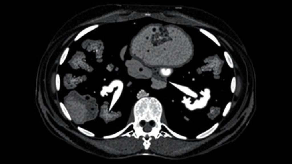
Adenocarcinoma of the esophagogastric junction, a cancer that arises where the esophagus meets the stomach, remains one of the most challenging malignancies to manage. Even after curative surgical resection, a substantial proportion of patients experience disease recurrence, and clinicians have long struggled to identify, before the operation, who is most likely to relapse. A new retrospective study published in BMC Medical Imaging by researchers at Lanzhou University Second Hospital in China suggests that two measurable features of a routine preoperative CT scan, when combined with standard clinical markers, can meaningfully sharpen the prediction of disease-free survival after surgery. The work, led by Xinyu Liu and Junlin Zhou, integrates spectral computed tomography-derived extracellular volume fraction with skeletal muscle index, offering a picture of both the tumor’s biology and the patient’s physical reserve from a single imaging examination.
The study enrolled 139 patients with adenocarcinoma of the esophagogastric junction who underwent curative resection. Of these, 65 patients experienced a disease-free survival event, meaning their cancer returned or they died during the follow-up period, while 74 patients remained free of disease. The researchers set out to determine whether imaging-derived measures could add predictive power beyond the conventional clinical factors that oncologists already use, such as tumor stage and serum tumor markers. To do this, they systematically analyzed clinical variables, body composition parameters, and spectral CT parameters, then used Cox regression analysis to identify which factors independently predicted worse disease-free survival.
Spectral CT, sometimes called dual-energy or energy-resolving CT, is a technology that distinguishes tissues not only by their conventional X-ray attenuation but also by how they behave at different X-ray energy levels. One of its most useful quantitative outputs is the extracellular volume fraction, a measure that reflects the proportion of extracellular space within a tissue. In tumors, an elevated extracellular volume fraction is generally interpreted as a sign of increased stromal content, tissue disorganization, and possibly more aggressive biology. Because spectral CT can quantify this property noninvasively from contrast-enhanced scans that patients already receive as part of standard staging, the researchers reasoned that it might serve as a preoperative window into tumor aggressiveness without requiring any additional invasive procedure.
The second pillar of the model is the skeletal muscle index, a body composition measure derived from the cross-sectional muscle area at a defined anatomical level, normalized for the patient’s height. Low skeletal muscle index, often described as sarcopenia, is a well-recognized marker of frailty and poor physiological reserve in cancer patients. In cancers of the upper gastrointestinal tract, where malnutrition and weight loss are common, muscle depletion has repeatedly been linked to worse surgical outcomes and survival. By quantifying muscle mass objectively from the same CT scan, the investigators could capture an aspect of the patient’s condition that is often missed by simple body weight or body mass index measurements.
The results of the Cox regression analysis identified four independent predictors of worse disease-free survival. Elevated carcinoembryonic antigen, or CEA, above 5 nanograms per milliliter carried the largest effect, with a hazard ratio of 3.31, indicating that patients with elevated CEA faced more than three times the hazard of a disease-free survival event compared with those below the threshold. Clinical T3 or T4 stage, reflecting deeper tumor invasion, more than doubled the hazard with a hazard ratio of 2.63. Low skeletal muscle index was associated with a hazard ratio of 2.18, and a high extracellular volume grade carried a hazard ratio of 1.98. All four associations reached statistical significance, with p values ranging from less than 0.001 to 0.026, confirming that both the tumor-derived spectral parameter and the patient-derived body composition measure contributed independent prognostic information.
To translate these findings into a practical tool, the researchers constructed three nested models of increasing complexity. Model 1 incorporated only the conventional clinical factors, CEA and clinical T stage. Model 2 added the body composition measure, the skeletal muscle index, and Model 3 further incorporated the spectral CT-derived extracellular volume grade. The models were assessed using time-dependent receiver operating characteristic analysis, which evaluates how well a model discriminates between patients who do and do not experience events at specific time points, along with calibration curves and decision curve analysis to judge clinical usefulness.
The performance gains from adding the imaging measures were consistent across all three time horizons evaluated. At one year, the area under the curve rose from 0.715 for Model 1 to 0.747 for Model 2 and 0.762 for Model 3. At two years, the corresponding values were 0.738, 0.759, and 0.773. At three years, the models achieved 0.768, 0.810, and 0.818 respectively. Although the incremental improvements in discrimination may appear modest in absolute terms, they were achieved using information that is already available before surgery, at no additional cost or risk to the patient. Calibration curves showed acceptable agreement between predicted and observed disease-free survival probabilities, and decision curve analysis supported the net clinical benefit of the fuller models across a range of threshold probabilities.
Perhaps the most striking finding emerged from an exploratory subgroup analysis that crossed the two imaging-derived variables. Patients who had both a high extracellular volume grade and a low skeletal muscle index showed the worst disease-free survival of any subgroup in the study. This combination is biologically plausible: it pairs a tumor that appears stroma-rich and potentially more aggressive on spectral imaging with a host whose muscle depletion suggests diminished reserve and possibly a more inflammatory, catabolic state. The convergence of adverse tumor biology and adverse host condition appears to compound risk in a way that neither measure captures alone, and the authors suggest this combined signature may support risk-adapted perioperative assessment and postoperative surveillance.
The practical implications of the study are considerable for the management of this tumor type. Adenocarcinoma of the esophagogastric junction sits at a diagnostic and therapeutic crossroads, treated variably by thoracic, upper gastrointestinal, and surgical oncology teams, and its prognosis after resection varies widely. A preoperative model built entirely from data available at staging, the CT scan and routine blood work, could help clinicians decide which patients warrant intensified neoadjuvant treatment, closer postoperative imaging surveillance, or earlier nutritional and physical interventions to address sarcopenia before it compounds recurrence risk. Because the model is preoperative, it does not depend on pathological examination of the resected specimen, which is only available after the operation when many treatment decisions have already been made.
The authors and readers alike should note the limitations inherent in the study design. It was a single-center retrospective analysis of 139 patients from Lanzhou University Second Hospital, and the model has so far been internally validated rather than tested on an independent external cohort. The exploratory subgroup findings, while compelling, require confirmation in larger and more diverse populations before they can change practice. The study was approved by the Institutional Review Board of the Second Hospital of Lanzhou University, with all clinical and imaging data fully anonymized before analysis, and the authors declare no competing interests. The research was supported by several Chinese funding bodies, including the National Natural Science Foundation of China and provincial science and technology programs. Even with these caveats, the study represents a concrete step toward a future in which the standard staging CT scan, already performed on nearly every patient, yields far more than tumor size and location, providing quantitative, reproducible measures of both the tumor’s microenvironment and the patient’s physical condition that together refine the forecast for life after cancer surgery.
Subject of Research: Preoperative spectral CT imaging biomarkers for predicting disease-free survival in esophagogastric junction adenocarcinoma
Article Title: Preoperative integration of spectral CT-derived extracellular volume and skeletal muscle index for disease-free survival prediction in adenocarcinoma of the esophagogastric junction
Article References: Liu, X., You, H., Ren, T., Chen, Q., Xu, M., Ma, L., Peng, Y., Zhang, C., Bao, J., Zhang, W., & Zhou, J. (2026). Preoperative integration of spectral CT-derived extracellular volume and skeletal muscle index for disease-free survival prediction in adenocarcinoma of the esophagogastric junction. BMC Medical Imaging. https://doi.org/10.1186/s12880-026-02873-4
Image Credits: AI Generated
DOI: 10.1186/s12880-026-02873-4
Keywords: esophagogastric junction adenocarcinoma, spectral CT, extracellular volume fraction, skeletal muscle index, sarcopenia, disease-free survival, prognosis, medical imaging, body composition, CEA, cancer staging, Cox regression
Cite Scienmag News
Ophelia Keating. (October 5, 2026). Spectral CT Muscle and Tissue Measures Predict Survival After Junctional Cancer Surgery. Scienmag. https://scienmag.com/spectral-ct-muscle-and-tissue-measures-predict-survival-after-junctional-cancer-surgery/
Ophelia Keating. "Spectral CT Muscle and Tissue Measures Predict Survival After Junctional Cancer Surgery." Scienmag, 5 October 2026, https://scienmag.com/spectral-ct-muscle-and-tissue-measures-predict-survival-after-junctional-cancer-surgery/. Accessed 5 October 2026.
Ophelia Keating. "Spectral CT Muscle and Tissue Measures Predict Survival After Junctional Cancer Surgery." Scienmag. October 5, 2026. https://scienmag.com/spectral-ct-muscle-and-tissue-measures-predict-survival-after-junctional-cancer-surgery/


