


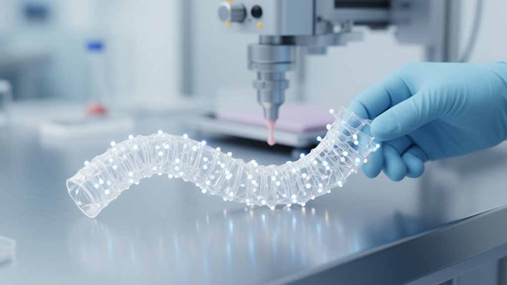
The trachea is one of the most deceptively demanding structures in the human body. It must hold an airway permanently open while flexing with every breath, cough, swallow and turn of the head, and its inner lining must continuously sweep mucus and trapped microbes upward and out of the lungs. When this tube is damaged by congenital malformations, trauma, prolonged intubation, infections or cancer, reconstruction remains one of the hardest problems in regenerative medicine. A new narrative review published in Frontiers in Sensors argues that the path forward may lie in combining 3D bioprinting with embedded biosensors, artificial intelligence and digital twin models to create tracheal grafts that can report on their own condition as they mature.
The review, authored by Sudipto Datta, Ranjit Barua, Saumya Dash and Sujata Mohanty from the All India Institute of Medical Sciences in New Delhi and OmDayal Group of Institution, surveys the state of tracheal tissue engineering from biomaterials and printing techniques to cell sources, vascularization strategies and the regulatory hurdles that stand between laboratory prototypes and approved implants. Its central thesis is that the field is shifting from static structural replacement toward what the authors call smart, sensor-assisted biological reconstruction of the airway.
Current clinical options all fall short in characteristic ways, the authors write. Autologous grafts are constrained by limited donor tissue and difficulty matching the native trachea’s mechanical and geometric properties. Allografts, though more readily available, carry risks of immune rejection, poor revascularization and long-term structural instability. Synthetic prostheses made of silicone or other polymers can provide temporary support but are prone to infection, migration and stenosis, and they do not integrate biologically with surrounding tissue. Earlier fabrication approaches have similar limits: electrospinning produces extracellular-matrix-like nanofibrous structures but cannot achieve the precise cylindrical geometry with graded mechanical properties; molding and casting give coarse shape control but homogeneous mechanics; and decellularized tracheal scaffolds, despite excellent biocompatibility, are difficult to strip of cells consistently and often lose structural integrity.
What makes the trachea so difficult to engineer is its heterogeneity. The human airway consists of 15 to 22 C-shaped cartilage rings joined by a flexible membranous posterior wall containing the trachealis muscle, creating spatial variations in stiffness that respond differently to breathing, coughing and head motion. The inner surface is lined with columnar epithelial cells that perform mucociliary clearance, chemoreception and antimicrobial defense, supported by a lamina rich in fibroblasts, immune cells, mucous glands, collagen and elastin. A functional graft must therefore reproduce not just the tubular shape but the anisotropic mechanics, the differentiated and polarized epithelium, and the segmental blood supply derived from the inferior thyroid, bronchial and subclavian arteries, a network the authors describe as fragmentary and easily damaged during surgery.
Three main cell types anchor the regeneration effort. Epithelial cells, including human primary tracheal epithelial cells, epithelial progenitors and airway basal stem cells, must rapidly form a contiguous barrier that prevents infection and mucus accumulation. Chondrocytes, whether harvested from tracheal or auricular cartilage or derived from mesenchymal stem cells stimulated with TGF-beta or BMP signaling, build the cartilage rings that prevent airway collapse. Fibroblasts produce the connective tissue matrix, while endothelial cells seed the microvascular networks a graft needs to survive. The review highlights induced pluripotent stem cells as a route to patient-specific, less immunogenic cell lines, though it cautions that cell line quality, scalability and system complexity remain unresolved problems.
On the fabrication side, extrusion-based bioprinting remains the most common technique for tracheal applications because it handles cell-laden hydrogels such as gelatin methacrylate, alginate, collagen and hyaluronic acid, and can incorporate stiffer polymers like polycaprolactone and PLGA for cartilage-mimetic rings. Its drawbacks are low printing resolution and high shear stress on cells. Inkjet bioprinting, ejecting picoliter droplets thermally or piezoelectrically, excels at depositing thin epithelial layers with minimal cell damage but struggles with viscous bioinks. Stereolithography and digital light processing achieve micro-level resolution ideal for external frameworks and cartilage-like rings using photopolymerizable bioresins such as PEGDA and GelMA. The authors conclude that no single method suffices: a functional tracheal substitute will likely require hybrid printing that combines extrusion, inkjet and light-based approaches, layered with electrospun fibers or SLA-printed frameworks.
The genuinely novel element of the review is its treatment of biosensing. Flexible electrochemical, optical and piezoelectric sensors can, in principle, be embedded into printed hydrogels to continuously track oxygen concentration, pH, glucose uptake, lactate production, temperature, mechanical strain and inflammatory markers. Such data would reveal hypoxia, infection or scaffold degradation early, and when coupled with wireless transmission and AI analytics could support predictive monitoring and adaptive control of bioreactor conditions. The authors are careful, however, to draw a firm line between what exists and what is aspiration: multifunctional sensors in fully printed tracheal grafts remain experimental, and capabilities such as hypoxia detection, infection tracking and AI-based evaluation in this specific application must be regarded as emerging or potential technologies rather than established features of clinically proven substitutes.
Conductive materials illustrate the same caution. Graphene derivatives, carbon nanotubes, MXenes, gold nanoparticles and conductive polymers such as PEDOT attract attention for biosensing and electrophysiological stimulation, but the review stresses that their biocompatibility cannot be assumed from physicochemical properties alone. Biological reactivity varies with composition, concentration, size, surface chemistry, functionalization, dispersion in the bioink, degradation products and exposure duration, so each formulation would require dedicated cytocompatibility, inflammation and stability testing. Responsive bioinks offer a complementary direction: hydrogels that release anti-inflammatory drugs under acidic conditions, or oxygen-sensitive materials that trigger angiogenic factor release in hypoxic regions, could turn static scaffolds into dynamic systems that react to the tissue environment.
Preclinical evidence is accumulating but uneven. The review tabulates animal studies in rabbits, dogs, pigs and mice, including 3D-printed polycaprolactone-plus-ECM grafts that achieved long-segment tracheal reconstruction with mucosal coverage and vascularized incorporation in pigs, biomimetic C-shaped constructs regenerated in rabbits and nude mice, and multilayered PCL-hydrogel implants seeded with nasal epithelial and auricular cartilage cells that maintained airway patency in rabbits, though cartilage regeneration remained limited. Chondro-fibrous integrated constructs using iPSC-derived chondrocytes and fibroblasts achieved segmental repair without collapse in rabbits and mice. Clinical experience remains sparse, and the authors note that inconsistent long-term outcomes, vascularization failures, immune reactions and the absence of standardized protocols still separate these results from routine clinical use.
The translational barriers the review catalogs are formidable. Sensors must survive cyclic mechanical loading, mucus exposure and sterilization without signal drift or adverse tissue reactions. Biodegradable, self-powered sensors that vanish after completing their monitoring role are proposed as a future direction, but they demand precise control over degradation timing, electrical stability and toxicity of breakdown products. Regulatory agencies would need to assess an unprecedented hybrid of living cells, degradable biomaterials, conductive elements, wireless electronics and software, spanning biological, mechanical, electrical and cybersecurity risks. Manufacturing would have to move from research-scale variability to GMP-compatible workflows with validated storage and transport. The authors frame AI and digital twins as computational aids for combining sensor modalities, optimizing print parameters and predicting tissue maturation, while emphasizing that this remains an emerging research field rather than a clinically adopted solution. Their overall verdict is measured: the integration of bioprinting, biosensing and digital health technologies may bring smart tracheal grafts closer to reality, but proof-of-concept demonstrations alone will not be enough to carry them into the clinic.
Subject of Research: A narrative review of 3D bioprinting, biosensing and digital technologies for engineering tracheal replacement grafts
Article Title: Translational advances in smart sensor-integrated 3D bioprinting for tracheal tissue engineering
Article References: Datta, S., Barua, R., Dash, S., & Mohanty, S. (2026). Translational advances in smart sensor-integrated 3D bioprinting for tracheal tissue engineering. Frontiers in Sensors, 7, Article 1961447. https://doi.org/10.3389/fsens.2026.1961447
Image Credits: AI Generated
DOI: 10.3389/fsens.2026.1961447
Keywords: 3D bioprinting, tracheal tissue engineering, biosensors, airway reconstruction, bioinks, regenerative medicine, digital twin, artificial intelligence, vascularization, biomaterials, preclinical studies, stem cells
Cite Scienmag News
Gregory Coleman. (October 27, 2026). Smart Biosensors and 3D Bioprinting Move Engineered Tracheas Closer to the Clinic. Scienmag. https://scienmag.com/smart-biosensors-and-3d-bioprinting-move-engineered-tracheas-closer-to-the-clinic/
Gregory Coleman. "Smart Biosensors and 3D Bioprinting Move Engineered Tracheas Closer to the Clinic." Scienmag, 27 October 2026, https://scienmag.com/smart-biosensors-and-3d-bioprinting-move-engineered-tracheas-closer-to-the-clinic/. Accessed 27 October 2026.
Gregory Coleman. "Smart Biosensors and 3D Bioprinting Move Engineered Tracheas Closer to the Clinic." Scienmag. October 27, 2026. https://scienmag.com/smart-biosensors-and-3d-bioprinting-move-engineered-tracheas-closer-to-the-clinic/


