


For men with advanced prostate cancer, blocking testosterone is one of the most powerful weapons in modern oncology. Androgen deprivation therapy, or ADT, starves tumors of the hormone that drives their growth, and it has become a cornerstone of treatment for locally advanced, node-positive, and metastatic disease. Yet the same hormonal suppression that constrains the cancer also reshapes the entire body, quietly dismantling muscle, expanding fat stores, and eroding the skeleton. A new study from Italian researchers now shows that the evidence of this body-wide transformation has been sitting inside patients’ medical records all along, embedded in the routine CT scans they already receive for cancer care.
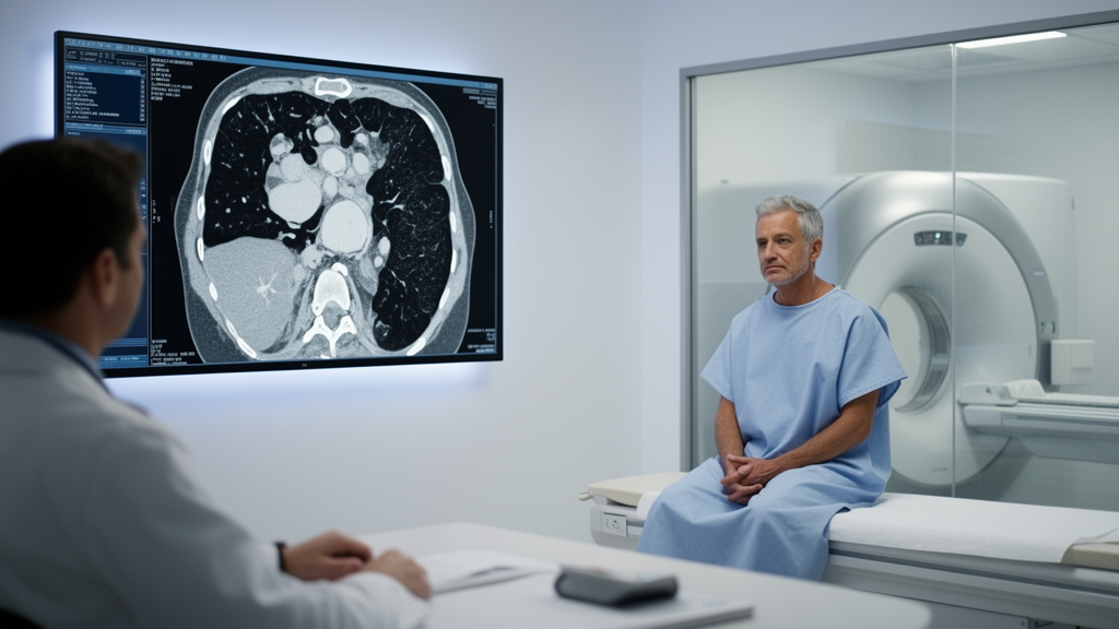
The research, published in Frontiers in Urology by a team led by Manuela Montatore and Giuseppe Guglielmi across the Universities of Foggia and hospitals in Brindisi, Bisceglie, and Barletti, examined 40 men who started ADT at Antonio Perrino Hospital in Brindisi between January 2023 and May 2025. Every patient had a contrast-enhanced CT scan within 30 days before treatment began and a technically comparable follow-up scan roughly a year later. Instead of ordering new imaging, the team extracted quantitative body-composition data from these existing oncologic examinations, an approach known as opportunistic imaging. The results paint a striking picture of simultaneous deterioration across three tissue compartments that conventional clinical measures largely missed.
The technical logic of the study rests on a well-validated principle of quantitative radiology: a single axial CT section at the third lumbar vertebra (L3) serves as a reliable surrogate for whole-body tissue composition. Using semiautomated threshold-based segmentation with attenuation windows of −29 to +150 Hounsfield units for muscle, −190 to −30 HU for subcutaneous fat, and −150 to −50 HU for visceral fat, the researchers measured the skeletal muscle index (SMI), bilateral psoas muscle attenuation, the subcutaneous adipose tissue index (SATI), and the visceral adipose tissue index (VATI). They then extended the analysis into the skeleton itself, placing oval regions of interest within the trabecular bone of the L1 to L3 vertebral bodies to capture vertebral attenuation, an opportunistic marker of bone health that no prior longitudinal ADT study has combined with muscle and fat in the same imaging framework.
After approximately twelve months of androgen suppression, the changes were substantial and consistent. Mean skeletal muscle index fell from 48.4 to 45.5 cm²/m², a relative decline of 6.0 percent. Mean psoas attenuation, which reflects the radiodensity of muscle tissue and indirectly captures fatty infiltration, dropped from 41.0 to 37.8 HU, a 7.8 percent reduction. Subcutaneous fat increased by 11.6 percent and visceral fat by 6.2 percent, while mean vertebral trabecular attenuation declined by 4.4 percent, from 132.1 to 126.3 HU, suggesting concurrent loss of bone quality. Twenty-three of the forty patients, or 57.5 percent, lost at least 5 percent of their skeletal muscle index, a prespecified exploratory threshold. All of these changes reached statistical significance, with p-values ranging from 0.018 to below 0.001.
What makes these findings especially compelling is what did not change: body mass index. BMI rose only modestly, from 26.7 to 27.0 kg/m², a difference that was not statistically significant, and the correlation between BMI change and SMI change was weak and nonsignificant (Spearman ρ = 0.24). The most telling statistic concerns the 23 men with clinically meaningful muscle loss: fifteen of them, or 65.2 percent, had a stable or increased BMI over the same period. Fat gain had effectively masked muscle depletion on the scale. This adiposity-driven camouflage means that a clinician relying on weight or BMI alone could wrongly conclude that a patient is tolerating treatment well, even as his metabolic reserve and physical resilience erode beneath a stable surface.
The study also explored whether any baseline feature could predict who would suffer the steepest muscle decline. In a deliberately parsimonious three-variable logistic regression model restricted to age, baseline psoas attenuation, and metastatic status, lower baseline psoas attenuation emerged as the only factor associated with a 5 percent or greater SMI decline, with an odds ratio of 1.14 per 1-HU decrease. Patients who ultimately lost the most muscle had started with noticeably darker, more fat-infiltrated muscle on their initial scans, averaging 38.9 HU compared with 43.8 HU in those with smaller declines. The authors are careful to frame this as hypothesis-generating: with only 40 patients, the model cannot establish psoas attenuation as an independent predictive biomarker, but it raises the possibility that a single number extracted from a pre-treatment scan could one day flag men at highest risk before therapy begins.
Why should androgen deprivation remodel muscle, fat, and bone simultaneously? Testosterone is an anabolic hormone that supports protein synthesis in skeletal muscle and suppresses adipogenesis. When it is pharmacologically eliminated, muscle catabolism accelerates, fat accumulates preferentially in visceral depots, and the delicate coupling between muscle and skeleton weakens. These compartments are not independent silos; they communicate through mechanical loading, hormonal signaling, inflammatory pathways, and metabolic crosstalk. A growing body of evidence links these tissue changes to hard clinical outcomes. Previous work has associated skeletal muscle loss during ADT with increased non-cancer mortality in high-risk prostate cancer, and more recent longitudinal studies have connected ADT-induced muscle loss and fat gain to subsequent cardiovascular events, suggesting that body composition may be a barometer of whole-patient vulnerability beyond oncologic status alone.
The reproducibility data strengthen the case that these measurements are robust rather than artifacts of measurement technique. When a second blinded radiologist independently re-analyzed ten patients’ paired scans, interobserver agreement was excellent: intraclass correlation coefficients reached 0.97 for SMI, 0.94 for psoas attenuation, 0.98 for SATI, 0.97 for VATI, and 0.93 for vertebral trabecular attenuation. The team also enforced strict technical comparability, requiring all paired scans to use the same portal venous contrast phase, matched tube voltage (120 kVp in 87.5 percent of pairs), and similar reconstruction thickness, because enhancement phase and acquisition parameters can shift attenuation values and contaminate longitudinal comparisons. Vertebral analysis proved feasible in 92.5 percent of potentially assessable vertebral levels, with metastatic involvement, compression deformities, or artifacts accounting for the few exclusions.
Important caveats temper the enthusiasm. The study was retrospective, single-center, and small, and it lacked an untreated comparator group, so the observed remodeling cannot be attributed exclusively to ADT. Aging, cancer progression, chemotherapy, radiotherapy, androgen-receptor pathway inhibitors, comorbidities such as diabetes and cardiovascular disease, and lifestyle factors may all have contributed. The five deaths recorded during follow-up were far too few for any survival inference, and the vertebral attenuation values from contrast-enhanced scans were explicitly not treated as DXA-equivalent bone mineral density or as diagnostic of osteoporosis. Patients who died before the follow-up scan could not contribute to the paired analysis, introducing potential survival bias. The authors emphasize that the study demonstrates feasibility, not proven clinical benefit, and that no treatment decisions should yet be triggered by these measurements.
Even so, the implications are provocative. CT scans are among the most common examinations in oncology, acquired routinely for staging, surveillance, and treatment assessment, and every one of them contains quantitative information about muscle, fat, and bone that currently goes unused. If larger, prospective, controlled studies confirm that integrated CT-derived phenotyping predicts fractures, metabolic complications, cardiovascular risk, or survival during ADT, then the humble staging scan could evolve into a longitudinal body-composition monitoring tool requiring no additional radiation, cost, or patient burden. For the millions of men worldwide who depend on androgen suppression to keep their cancer in check, the study suggests a future in which the price of treatment is measured not just in tumor control, but in the health of every tissue it touches, visible in images doctors already possess.
Subject of Research: CT-derived body composition changes during androgen deprivation therapy for prostate cancer
Article Title: Longitudinal CT-derived muscle–fat–bone remodeling following androgen deprivation therapy initiation in prostate cancer
Article References: Montatore, M., Muscatella, G., Masino, F., Muscogiuri, E., & Guglielmi, G. (2026). Longitudinal CT-derived muscle–fat–bone remodeling following androgen deprivation therapy initiation in prostate cancer. Frontiers in Urology, 6, Article 1985221. https://doi.org/10.3389/fruro.2026.1985221
Image Credits: AI Generated
DOI: 10.3389/fruro.2026.1985221
Keywords: androgen deprivation therapy, prostate cancer, computed tomography, body composition, skeletal muscle index, sarcopenia, visceral adiposity, vertebral trabecular attenuation, opportunistic imaging, bone health, BMI, oncology
Cite Scienmag News
Nathaniel Bowman. (October 15, 2026). Routine CT Scans Reveal Hidden Muscle Loss, Fat Gain, and Bone Thinning in Prostate Cancer Patients on Hormone Therapy. Scienmag. https://scienmag.com/routine-ct-scans-reveal-hidden-muscle-loss-fat-gain-and-bone-thinning-in-prostate-cancer-patients-on-hormone-therapy/
Nathaniel Bowman. "Routine CT Scans Reveal Hidden Muscle Loss, Fat Gain, and Bone Thinning in Prostate Cancer Patients on Hormone Therapy." Scienmag, 15 October 2026, https://scienmag.com/routine-ct-scans-reveal-hidden-muscle-loss-fat-gain-and-bone-thinning-in-prostate-cancer-patients-on-hormone-therapy/. Accessed 15 October 2026.
Nathaniel Bowman. "Routine CT Scans Reveal Hidden Muscle Loss, Fat Gain, and Bone Thinning in Prostate Cancer Patients on Hormone Therapy." Scienmag. October 15, 2026. https://scienmag.com/routine-ct-scans-reveal-hidden-muscle-loss-fat-gain-and-bone-thinning-in-prostate-cancer-patients-on-hormone-therapy/


