Researchers have built a collagen-based hydrogel that combines an ordered internal architecture with on-demand drug delivery triggered by near-infrared light, and laboratory tests show it can steer fibroblasts deep into the material while calming inflammatory signaling in immune cells. The material, described in the journal Collagen and Leather, was developed by a team led by Hui Chen, Defu Li and Liming Ge and is designed around two persistent bottlenecks in tissue repair: cells that fail to infiltrate a wound scaffold efficiently, and inflammation that lingers long after injury.
The hydrogel, named DOCH@P, is assembled from type I collagen extracted from bovine Achilles tendon and oxidized hyaluronic acid, which crosslink through Schiff base chemistry. Embedded within this matrix are nanoparticles that carry the cargo that gives the material much of its biological activity. Mesoporous polydopamine nanoparticles, roughly 159 nanometers in diameter with an average pore size of 21.0 nanometers, were loaded with spermidine, a natural polyamine known for immunomodulatory effects, and then coated with an agarose shell about 8 nanometers thick. The coating raised the particle diameter to 175 nanometers and, crucially, sealed the spermidine inside until heat unlocked it.
That lock-and-key mechanism is physical rather than chemical. Under irradiation from an 808-nanometer near-infrared laser at a power density of 1.5 watts per square centimeter, the polydopamine cores convert light into heat through non-radiative relaxation of their conjugated pi-electron system. The agarose shell melts, surface pores reappear, and the spermidine escapes. Without light, the coated nanoparticles released almost none of their payload, only a small fraction diffusing from the outer layer. With light, release was rapid and followed a sigmoidal profile that the authors fitted best with a Logistic kinetic model. In the finished hydrogel, only about 3.9 percent of the spermidine leaked out in the dark, while cumulative release reached 65.2 percent within eight hours after irradiation.
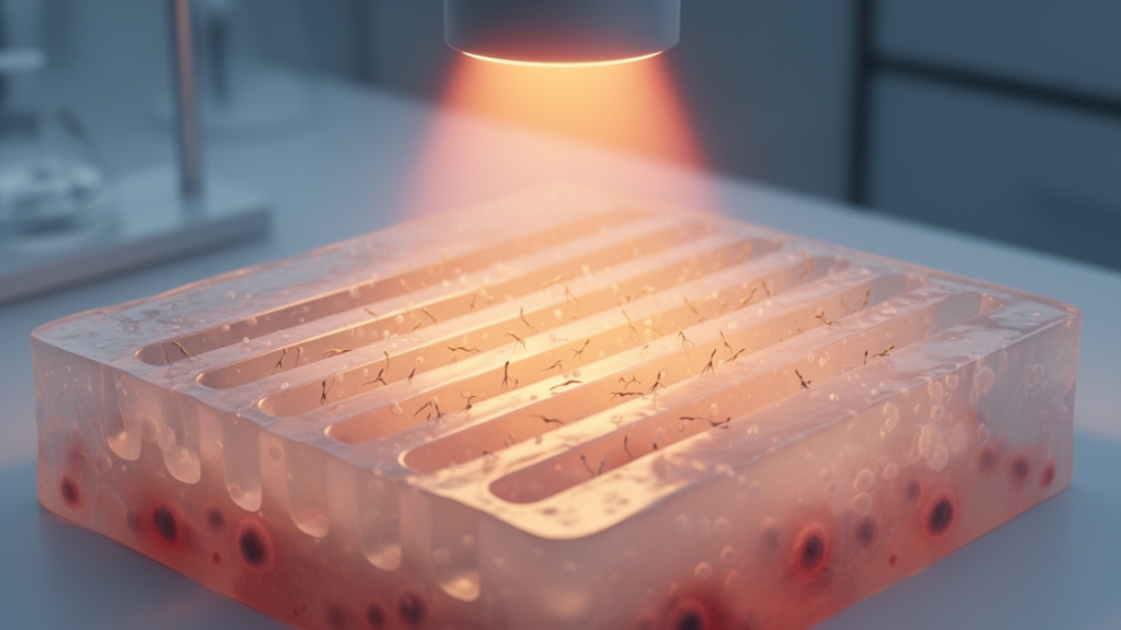
The hydrogel itself was shaped by a directional freeze-thaw process. The precursor mixture was poured into a mold and frozen from the bottom with liquid nitrogen for five minutes, forcing ice crystals to grow upward and squeeze the polymer network into parallel channels. During a subsequent 14-day hold at minus 20 degrees Celsius, the Schiff base crosslinking between collagen and oxidized hyaluronic acid continued, locking the aligned architecture in place so that it survived thawing at 37 degrees. Scanning electron microscopy confirmed distinct oriented microchannels in the directionally frozen gels, while an unfrozen control showed only an irregular porous structure. The channel structure remained intact after two days in simulated body fluid, though it partially collapsed after five days.
The aligned architecture also paid mechanical dividends. DOCH@P reached a maximum compressive strength of 34.7 kilopascals and a Young’s modulus of 4.3 kilopascals, values the authors attribute to the dense, load-bearing channel array and to non-covalent interactions between the nanoparticles and the collagen network. Rheology testing showed the gel-sol transition point shifted to a strain of 25.2 percent, well above the 9.6 and 10.1 percent measured for the two control gels, indicating greater viscoelastic stability. The material also adhered to fresh pigskin with a strength of 12.0 kilopascals, roughly double the 5.0 to 5.2 kilopascals recorded for the controls, an effect credited to hydrogen bonding from the agarose coating and the nanoparticle surface groups.
Photothermal heating gave the hydrogel a second function: killing bacteria. Irradiated DOCH@P reached 60.9 degrees Celsius within five minutes, while gels lacking the nanoparticles showed no meaningful temperature change. In co-culture assays with Escherichia coli and Staphylococcus aureus, representatives of gram-negative and gram-positive bacteria, the irradiated gel achieved inhibition efficiencies exceeding 95 percent against both species. Microscopy revealed roughened, shrunken and ruptured bacterial membranes in the irradiated group, consistent with heat denaturation of bacterial proteins and light-triggered oxidative damage. The authors note that photothermal killing, unlike antibiotics, does not select for resistance, and the non-irradiated gels showed no significant antibacterial effect.
The material also scavenged reactive chemical species in vitro. In ABTS radical cation and hydroxyl radical assays, the irradiated hydrogel outperformed all controls, an effect the researchers attribute to the synergy of phenolic hydroxyl and quinone groups on the now-exposed polydopamine cores, which trap radicals through electron transfer, and the proton-donating amino groups of the newly released spermidine. Because the photothermal treatment itself can generate oxidative stress in surrounding tissue, the authors frame this antioxidant activity as a way of offsetting the collateral chemistry of its own antibacterial mechanism.
Cell experiments pointed to the scaffold’s central promise. L929 fibroblasts seeded on the hydrogels survived at rates above 85 percent with low cytotoxicity grades of 0 to 1, and the migration rate in the irradiated DOCH@P group exceeded 80 percent within three days, the best of any formulation. Confocal microscopy showed cells aligning, migrating directionally and infiltrating deeply along the microchannels in the directionally frozen gels, while cells on the unfrozen control infiltrated in a disordered fashion. The authors attribute this to the combination of physical contact guidance from the aligned channels and spermidine-driven cytoskeletal reorganization, including F-actin polymerization and enhanced pseudopod formation. Blood compatibility tests showed low clotting indices and hemolysis rates below the 5 percent threshold generally required of biomedical scaffolds.
The immunological results came from macrophage experiments rather than animal or human tissue. When lipopolysaccharide-stimulated RAW264.7 macrophages were treated with the irradiated hydrogel, protein and mRNA levels of the pro-inflammatory cytokines IL-1beta, IL-6 and TNF-alpha and the M1 marker i-NOS fell, while the anti-inflammatory cytokine IL-10 and the M2 markers Arg-1 and CD206 rose, alongside reduced NF-kappa-B expression. Transcriptome sequencing of the same cells identified 3,190 differentially expressed genes, 846 up-regulated and 2,344 down-regulated. Down-regulated genes clustered in TNF, NF-kappa-B, IL-17 and PI3K-AKT signaling pathways, while up-regulated genes enriched in autophagy-related PPAR and mTOR pathways and in cytochrome P450 and glutathione metabolism, a pattern the authors interpret as spermidine simultaneously suppressing inflammatory signaling and promoting cellular cleanup through autophagy.
The authors conclude that the light-activated, channel-structured collagen hydrogel offers a scaffold foundation for guiding tissue regeneration through contact-guided cellular infiltration and inflammation resolution, with potential application in chronic skin wound repair. As the evidence stands, however, every functional result comes from in vitro assays with bacterial cultures, fibroblast cell lines and macrophage cell lines; no animal wound model or human data appear in the reported work.
Subject of Research: A near-infrared-activated collagen hydrogel with oriented microchannels for controlled spermidine release, antibacterial activity and macrophage immunomodulation in tissue engineering.
Article Title: Near-infrared light-activated collagen-based hydrogel with oriented microchannel structure promoting fibroblast infiltration and inflammatory resolution
Article References: Chen, H., Xu, Y., Zhou, Y., Xu, Z., Guo, J., Mu, C., Li, D., & Ge, L. (2026). Near-infrared light-activated collagen-based hydrogel with oriented microchannel structure promoting fibroblast infiltration and inflammatory resolution. Collagen and Leather, 8(1), Article 35. https://doi.org/10.1186/s42825-026-00258-5
Image Credits: AI Generated
DOI: 10.1186/s42825-026-00258-5
Keywords: collagen hydrogel, near-infrared light, spermidine, polydopamine nanoparticles, tissue engineering, fibroblast migration, macrophage polarization, photothermal antibacterial, directional freeze-thaw, oxidized hyaluronic acid, anti-inflammatory, wound scaffold
Cite Scienmag News
Denise Maddox. (October 28, 2026). Research report: Near-infrared light-activated collagen-based hydrogel with oriented microchannel structure promoting fibroblast infiltration and inflammatory resolution. Scienmag. https://scienmag.com/research-report-near-infrared-light-activated-collagen-based-hydrogel-with-oriented-microchannel-structure-promoting-fibroblast-infiltration-and-inflammatory-resolution/
Denise Maddox. "Research report: Near-infrared light-activated collagen-based hydrogel with oriented microchannel structure promoting fibroblast infiltration and inflammatory resolution." Scienmag, 28 October 2026, https://scienmag.com/research-report-near-infrared-light-activated-collagen-based-hydrogel-with-oriented-microchannel-structure-promoting-fibroblast-infiltration-and-inflammatory-resolution/. Accessed 29 October 2026.
Denise Maddox. "Research report: Near-infrared light-activated collagen-based hydrogel with oriented microchannel structure promoting fibroblast infiltration and inflammatory resolution." Scienmag. October 28, 2026. https://scienmag.com/research-report-near-infrared-light-activated-collagen-based-hydrogel-with-oriented-microchannel-structure-promoting-fibroblast-infiltration-and-inflammatory-resolution/