Micro-computed tomography and bone-by-bone segmentation have corrected several interpretations of the skull of Allaeochelys libyca , a Middle Miocene carettochelyid turtle from Libya and the only valid African taxon currently recognized in its family. The digital redescription confirms the species’ validity while revealing previously inaccessible details of its cranial nerves, blood vessels, inner ear and skull bones. Because the fossil cranium is incomplete, some anatomical reconstructions remain inferential, but the study identifies structures that could contribute to finer evolutionary comparisons among carettochelyids.
The specimen examined is BSPG 1991 II 130, the holotype of Allaeochelys libyca , held by the Bayerische Staatssammlung für Paläontologie und Geologie in Munich. It is an incomplete cranium from the Lower Maradah Formation at Gebel Zelten, also called Jabal Zaltan, in southwestern Libya. The locality has been assigned to the Langhian stage of the Middle Miocene.
The species was originally described in 2014 from this partial skull and several isolated shell fragments. That earlier work described the externally visible anatomy, but the internal cavities, canals and other structures could not be examined directly. The new reassessment began when the researchers downloaded the specimen’s micro-CT slice data from the MorphoSource repository and noticed that their three-dimensional reconstructions differed from the original interpretation of some features.
The fossil had been scanned at the University of Bristol’s X-ray Tomography Facility using a Nikon Metrology XT H 225 ST scanner at 125 kV and 265 µA. The scan produced 1,601 projections and 1,813 coronal slices with a voxel size of 37.8 µm. The researchers segmented individual bones in Mimics Innovation Suite 25, exported the objects as PLY files and visualized them in Blender 2.79b.
The resulting cranium and inner-ear models were also deposited at MorphoSource, making the digital reconstruction available for further study. One of the most consequential revisions concerns the opening that the original description called the “posterior” trigeminal nerve foramen. The CT data show that it communicates with the cavum acustico-jugulare, a chamber associated with the inner ear and major cranial nerves, rather than opening directly into the braincase in which the trigeminal nerve originates.
Comparisons with the living pig-nosed turtle, Carettochelys insculpta , indicate that the fossil opening represents the posterior portion of a broader passage associated with both the trigeminal nerve and the mandibular artery. The authors distinguish the true trigeminal foramen from the posterior mandibular artery foramen, while noting that the two are homologous with the combined “trigeminal foramen sensu lato” of the living species. Reconstructing the missing epipterygoid is essential to that interpretation.
Havlik and colleagues had identified a purported epipterygoid beneath the processus trochlearis oticum and between the two openings, but the CT slices show that the apparent sutures around this piece are actually a crack. The bone is not preserved. A small, anteroventrally projecting bump on the fossil’s prootic, however, matches the articular site for the epipterygoid in Carettochelys insculpta .
This supports the inference that the missing bone formed a narrow bridge around a strongly reduced canalis cavernosus. In the living turtle, that canal is closely associated with the lateral head vein, while the fossil suggests that the mandibular artery could pass from the inner-ear region into the lower temporal fossa. The authors consider complete loss of the canal in Allaeochelys followed by its reappearance in Carettochelys less likely than preservation of a reduced canal obscured by the missing epipterygoid.
The scans also clarify the circulatory routes through the skull. The posterior opening of the internal carotid canal lies entirely within the pterygoid, rather than in the more anteroventral position occupied by the corresponding foramen in the early-branching carettochelyids Anosteira pulchra and Anosteira maomingensis . From this opening, the internal carotid canal extends anteromedially through the pterygoid and continues into the parabasisphenoid as the basisphenoidal carotid canal.
A branch of the facial nerve for the vidian nerve joins the internal carotid canal, while a separate canal carries the abducens nerve through the parabasisphenoid. As in Carettochelys insculpta and Anosteira pulchra , no lateral carotid canal is present. The reconstructed arterial and nervous routes are therefore similar across these taxa, even though the position of the posterior carotid opening differs in A. libyca .
Neural anatomy also yielded a correction to the count of hypoglossal foramina. The original description recognized three external openings for the hypoglossal nerves, but tomography reveals only two distinct sets of internal and external foramina, together with their associated canals. The supposed third external opening is exposed surface porosity rather than a nerve passage.
The two-foramina condition matches the living Carettochelys insculpta and the fossil Anosteira pulchra . The reinterpretation also places the nearly horizontal suture between the exoccipital and basioccipital farther ventrally than in the original description, although the seam fades toward the posterior and was therefore difficult to follow without tomographic data. The endosseous labyrinth, the bony housing of the inner ear, is among the structures newly documented in detail.
Its semicircular canals are thick, with the anterior canal the longest and the lateral canal the shortest. The anterior canal joins the vestibule at the level of the anterior ampulla. The posterior canal is shorter, and its posterior third is confluent with the posterior part of the lateral canal, creating a large secondary common crus.
The lateral canal forms a proper canal only along its posterior half and merges anteriorly with a large lateral ampulla. The fossil labyrinth is extremely similar to that of the fully segmented Carettochelys insculpta specimen NHMUK 1903.7.10.1. Only two subtle differences were identified: the anterior semicircular canal of the fossil is slightly thicker, and the dorsal embayment of the common crus is slightly more deeply excavated.
Other findings refine the skull’s construction. A secondary contact between the frontal and prefrontal appears to create a passage between the orbit and nasal cavity, a condition also reported in the Middle Miocene Carettochelys niahensis but absent in the living species. The fossil also possesses an anterior quadrate foramen and a short canal that the authors name as a potential shared feature of derived carettochelyids; its precise identity remains uncertain because no connection with the columella auris or the cavum acustico-jugulare could be demonstrated.
A large recessus scalae tympani, a possible glossopharyngeal nerve canal in the opisthotic, and the arrangement of the jugular and hypoglossal foramina were also reconstructed. These observations remain constrained by missing premaxillae, maxillae, jugals, vomer, squamosals and other damaged elements, as well as the loss of much of the epipterygoid and anterior braincase. The anatomical revisions do not challenge Allaeochelys libyca as a valid species.
Its broader and shorter cranium, secondary prefrontal–frontal contact, deeper pterygoid fossa, larger quadrate fossa, reduced canalis cavernosus and other traits continue to distinguish it from Carettochelys insculpta . The authors do expect the redescription to affect phylogenetic matrices, where several character scorings will need revision and additional features may be added, including the prefrontal–frontal contact, the possible glossopharyngeal canal and the relative length of the basioccipital tubercles. Although these changes are not expected to produce a drastic rearrangement of carettochelyid relationships, they may help resolve subclades that have remained uncertain, including the groups containing Anosteira and Allaeochelys .
The study demonstrates how a single damaged fossil can preserve a detailed record of sensory, neural and vascular anatomy that was previously hidden inside a skull.
Subject of Research: Digital tomographic redescription of the Middle Miocene carettochelyid turtle Allaeochelys libyca
Article Title: A digital redescription of the Middle Miocene (Langhian) carettochelyid turtle Allaeochelys libyca
Article References: Rollot, Y., Evers, S. W., & Joyce, W. G. (2024). A digital redescription of the Middle Miocene (Langhian) carettochelyid turtle Allaeochelys libyca. Fossil Record, 27(1), 13-28. https://doi.org/10.3897/fr.27.115046
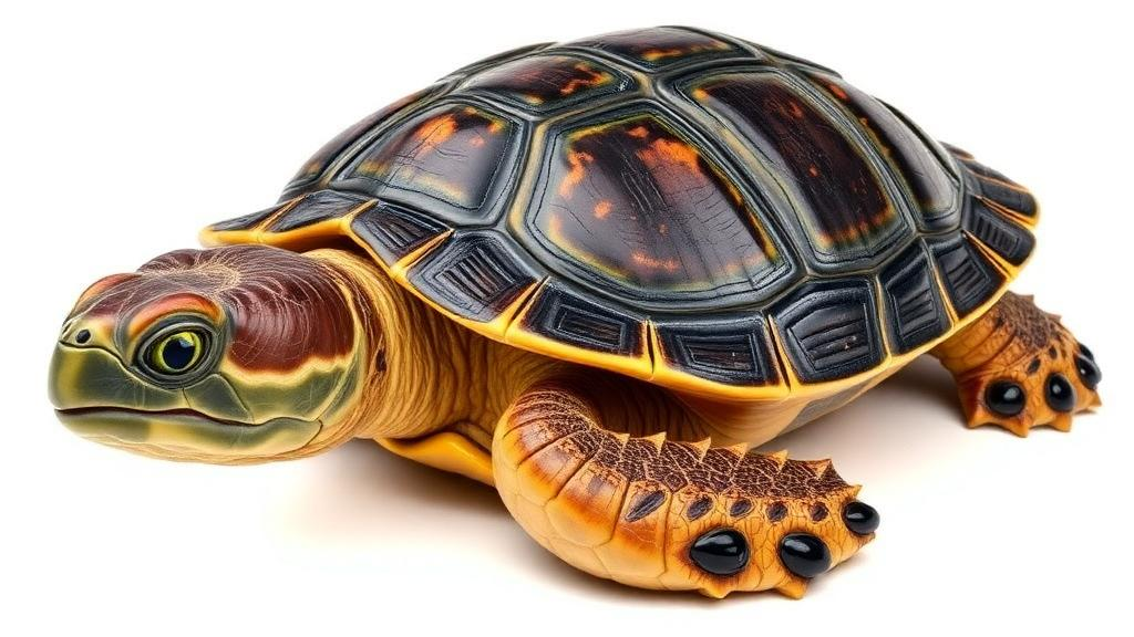
Image Credits: AI Generated
DOI: 10.3897/fr.27.115046
Keywords: Allaeochelys libyca, Carettochelyidae, micro-computed tomography, fossil skull anatomy, Miocene turtle, paleontology, inner ear, cranial nerves, digital reconstruction, Libya, digital, redescription
Cite Scienmag News
Gavin Prescott. (October 20, 2026). Research report: A digital redescription of the Middle Miocene (Langhian) carettochelyid turtle Allaeochelys libyca. Scienmag. https://scienmag.com/research-report-a-digital-redescription-of-the-middle-miocene-langhian-carettochelyid-turtle-allaeochelys-libyca/
Gavin Prescott. "Research report: A digital redescription of the Middle Miocene (Langhian) carettochelyid turtle Allaeochelys libyca." Scienmag, 20 October 2026, https://scienmag.com/research-report-a-digital-redescription-of-the-middle-miocene-langhian-carettochelyid-turtle-allaeochelys-libyca/. Accessed 20 October 2026.
Gavin Prescott. "Research report: A digital redescription of the Middle Miocene (Langhian) carettochelyid turtle Allaeochelys libyca." Scienmag. October 20, 2026. https://scienmag.com/research-report-a-digital-redescription-of-the-middle-miocene-langhian-carettochelyid-turtle-allaeochelys-libyca/