


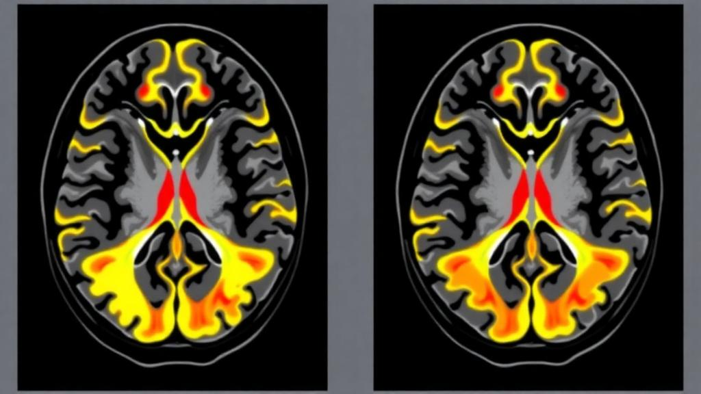
Sarcomas are among the rarest and most bewildering cancers in medicine. Arising not from organs but from connective tissues such as bone, cartilage, fat, muscle, and blood vessels, they encompass dozens of distinct subtypes, each with its own biology and behavior. When these tumors spread to the brain, clinicians have long operated in a fog: case reports exist, but systematic descriptions of what sarcoma metastases actually look like on brain scans have been almost entirely missing. A new study from a tertiary referral center in Padua, Italy, now offers one of the clearest pictures to date, and its findings suggest that some sarcoma subtypes may stamp a recognizable fingerprint on neuroimaging that radiologists could one day use to guide diagnosis and treatment.
The research, published as an open-access article in BMC Cancer, set out to systematically characterize the computed tomography (CT) and magnetic resonance imaging (MRI) features of intracranial sarcoma metastases. The team, led by neuroradiologist Giovanni Librizzi and oncologist Benedetta Chiusole, together with senior author Renzo Manara, reviewed the records of patients followed for intracranial sarcoma metastases between January 2011 and April 2024. To keep the analysis rigorous, they excluded patients who lacked pretreatment brain imaging and those whose disease was confined to the skull itself. What remained was a cohort of 22 patients whose pre-treatment scans could be scrutinized in detail, comprising 15 MRI studies and 18 CT examinations, spanning twelve different sarcoma tumor types.
The methodology was deliberately meticulous. Two neuroradiologists, blinded to the pathological diagnosis, independently assessed each patient’s imaging. They recorded the number and location of metastases, the size of the largest lesion, and a battery of morphological features: the presence of perilesional edema, calcification, hemorrhage, cysts, necrosis, and leptomeningeal disease, in which tumor cells spread through the membranes surrounding the brain. They also measured CT density and MRI signal intensity both before and after intravenous contrast administration, quantified Apparent Diffusion Coefficient values on diffusion-weighted imaging, and evaluated perfusion characteristics. This comprehensive approach meant that essentially every dimension of modern neuroradiological assessment was brought to bear on a disease entity that had never been systematically profiled in this way.
The headline finding is heterogeneity, and lots of it. Ten of the 22 patients presented with multiple brain metastases from the outset, and one patient had diffuse leptomeningeal seeding, meaning tumor cells had spread widely through the cerebrospinal fluid spaces. More striking still, the lesions were heterogeneous not only between patients with different tumor types but also between different metastases within the same patient. In practical terms, two tumors sitting in the same brain, originating from the same primary sarcoma, could look dramatically different on the same scan, differing in density, signal intensity, enhancement pattern, and surrounding swelling. For radiologists accustomed to the relatively predictable appearance of metastases from lung, breast, or melanoma primaries, this variability presents a genuine diagnostic challenge.
Yet within that chaos, the researchers identified patterns that appear to be associated with specific sarcoma subtypes. All three patients with chondrosarcoma metastases in the cohort displayed the T2-FLAIR mismatch sign, an imaging phenomenon in which a lesion appears bright on T2-weighted sequences but does not suppress, or darken, on FLAIR sequences. This sign is familiar to neuroradiologists as a hallmark of certain low-grade gliomas, particularly those with specific mutations, but its appearance in chondrosarcoma metastases had not been emphasized before. The sign was absent or uncommon in the other sarcoma subtypes studied, making it a candidate marker that could raise suspicion of chondrosarcoma when a patient with an unknown primary presents with brain lesions.
Ewing sarcoma, an aggressive tumor most often seen in children and young adults, showed an equally distinctive pattern. Every Ewing sarcoma patient in the study had exclusively pachymeningeal disease, meaning the tumor involved the tough outer dura mater rather than forming discrete masses within the brain parenchyma. This dural predilection distinguishes Ewing metastases from the intraparenchymal lesions typical of many other cancers and could help clinicians interpret scans in young patients with known or suspected Ewing sarcoma. Meanwhile, osteosarcoma patients showed yet another signature: their brain metastases exhibited gross calcification, visible as dense, bone-like deposits on CT. This feature makes intuitive biological sense, given that osteosarcoma is a tumor that produces osteoid, or bone matrix, but confirming it radiologically provides a useful diagnostic clue. These three features, the T2-FLAIR mismatch in chondrosarcoma, pachymeningeal disease in Ewing sarcoma, and calcification in osteosarcoma, were absent or uncommon in the other subtypes examined.
The study also documented genuinely unusual presentations that underline how deceptive these tumors can be. One chondrosarcoma patient harbored a mixture of conventional brain metastases and multiple neoplastic emboli, fragments of tumor that had lodged in cerebral blood vessels, producing a picture that could easily be mistaken for stroke or vascular disease. In another remarkable case, a leiomyosarcoma metastasis was invisible on standard sequences and could be detected only on diffusion-weighted imaging, the MRI technique that is sensitive to the random motion of water molecules and is typically used to identify acute stroke. Such cases serve as cautionary tales: a routine contrast-enhanced brain MRI protocol, interpreted without awareness of these possibilities, could theoretically miss disease or mischaracterize it entirely.
Why does this matter clinically? Brain metastases from sarcomas are rare, but their incidence may be rising as systemic therapies extend survival and allow more patients to live long enough for intracranial spread to manifest. Treatment decisions, whether surgery, stereotactic radiosurgery, whole-brain radiotherapy, or systemic agents, often hinge on the number, size, and location of lesions, and increasingly on the biological behavior inferred from imaging. If certain imaging features reliably point to a specific sarcoma subtype, they could accelerate diagnosis in patients whose primary tumor is unknown, inform biopsy planning, and help oncologists anticipate complications such as hemorrhage or leptomeningeal spread. The T2-FLAIR mismatch sign, in particular, is intriguing because it links sarcoma imaging to a well-established concept in neuro-oncology, potentially opening a bridge between two fields that rarely intersect.
The authors are appropriately cautious about overinterpreting their results. With 22 patients and twelve tumor types, the cohort is small, and several subtypes are represented by only one or two individuals. The candidate type-associated imaging features they identified require confirmation in larger, multicenter cohorts before they can be incorporated into diagnostic algorithms or clinical guidelines. Rare-disease research inevitably faces this tension: the very rarity that makes sarcoma metastases poorly understood also makes it difficult to assemble the patient numbers needed for statistically robust conclusions. Single-center studies like this one are best viewed as hypothesis-generating, laying the groundwork for collaborative efforts that pool cases across institutions and countries.
Even so, the study represents a meaningful step forward for a neglected corner of neuro-oncology. By applying a systematic, blinded, quantitative imaging analysis to a group of patients whose scans had never been collectively characterized, the Padua team has transformed scattered anecdotes into a structured spectrum. The message for radiologists and oncologists is twofold: first, assume nothing when interpreting brain lesions in sarcoma patients, because heterogeneity is the rule both within and between individuals; and second, pay attention to specific clues, FLAIR behavior, dural involvement, and calcification, that may whisper the tumor’s identity. As survival improves for sarcoma patients worldwide, the brain is becoming an increasingly important frontier in their care, and studies like this one ensure that the imaging tools needed to meet that challenge are finally beginning to take shape.
Subject of Research: Neuroradiological features of intracranial metastases from sarcoma subtypes
Article Title: Neuroradiological spectrum of sarcoma intracranial metastases: a single center study
Article References: Librizzi, G., Murtas, F., Chiusole, B., Tortorelli, I., Valeggia, S., Vizzaccaro, S., Di Maggio, A., Buzzaccarini, M. S., Ghirelli, A., Brunello, A., & Manara, R. (2026). Neuroradiological spectrum of sarcoma intracranial metastases: a single center study. BMC Cancer. https://doi.org/10.1186/s12885-026-17009-8
Image Credits: AI Generated
DOI: 10.1186/s12885-026-17009-8
Keywords: sarcoma, brain metastases, neuroradiology, MRI, CT, chondrosarcoma, Ewing sarcoma, osteosarcoma, T2-FLAIR mismatch, leptomeningeal disease, cancer imaging, tumor heterogeneity
Cite Scienmag News
Nathaniel Bowman. (October 11, 2026). Rare Sarcoma Brain Metastases Show Striking Imaging Signatures on MRI Scans. Scienmag. https://scienmag.com/rare-sarcoma-brain-metastases-show-striking-imaging-signatures-on-mri-scans/
Nathaniel Bowman. "Rare Sarcoma Brain Metastases Show Striking Imaging Signatures on MRI Scans." Scienmag, 11 October 2026, https://scienmag.com/rare-sarcoma-brain-metastases-show-striking-imaging-signatures-on-mri-scans/. Accessed 11 October 2026.
Nathaniel Bowman. "Rare Sarcoma Brain Metastases Show Striking Imaging Signatures on MRI Scans." Scienmag. October 11, 2026. https://scienmag.com/rare-sarcoma-brain-metastases-show-striking-imaging-signatures-on-mri-scans/


