


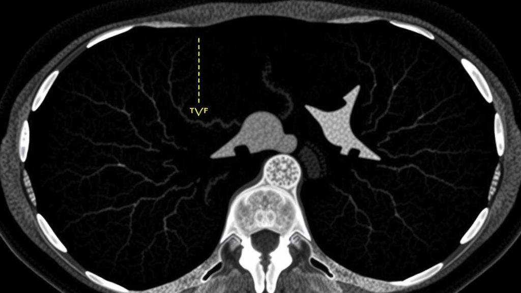
Forensic radiologists in Zurich have identified a strikingly rare imaging feature that could sharpen one of the most consequential judgments in gunshot death investigations: distinguishing an entry wound from an exit wound, and in some cases inferring how close the muzzle was to the skin when the trigger was pulled. In a retrospective analysis of fifty forensic cases published in the International Journal of Legal Medicine, researchers at the Institute of Forensic Medicine, University of Zurich report that a compact, well-demarcated ring of radiopaque material at the wound margin—dubbed the radiopaque ring sign—appeared in only three of fifty cases, or six percent of the cohort. All three of those cases, the team found, involved revolvers and external findings consistent with contact shots, suggesting the sign may be tied to very specific firing conditions rather than being a universal feature of gunshot residue deposition.
Postmortem computed tomography, or PMCT, has become a mainstay of modern forensic investigation, offering a non-invasive window into projectile trajectories, fracture patterns, and the extensive osseous destruction typical of cranial gunshot injuries. Established PMCT findings include inward and outward bevelling of bone, keyhole lesions, radiating fracture lines, and the distribution of bone fragments, air, and blood along the bullet path. Yet comparatively little systematic attention has been paid to the hyperdense—radiographically bright—findings that appear at the entry wound itself. These bright foci are frequently attributed to gunshot residue, the particulate and combustion-related material expelled from a firearm upon discharge, but their morphology and underlying mechanisms have remained poorly differentiated, despite their potential forensic weight.
The new study set out to determine whether the various hyperdense patterns seen at entry sites represent a single phenomenon or a heterogeneous spectrum. Led by Radiya Sri Rajendran, with Stephan Andreas Bolliger, Michael Josef Thali, and Dominic Gascho as co-authors, the team analyzed fifty cases of gunshot wounds to the head and neck, all considered consistent with short-range shots based on forensic documentation. Thirty-five cases, seventy percent, were through-and-through injuries with an exit wound, while fifteen cases, thirty percent, involved retained projectiles. Pistols accounted for sixty-six percent of the weapons, revolvers for twenty-six percent, and rifles for eight percent, with 9 mm Para and .22 long rifle ammunition the most frequently represented calibers.
Imaging was performed on a 128-slice SOMATOM Definition Flash scanner using standardized parameters: a tube voltage of 120 kV, a tube current-time product of 400 mAs, a pitch of 0.35, and a rotation time of 1.0 second. Datasets were reconstructed at a slice thickness of 0.6 millimeters with a bone kernel to optimize visualization of radiopaque structures. Two readers with very different levels of experience independently assessed gunshot residue-related hyperdensities: Reader 1 was a physician without specific training in forensic radiology or wound ballistics, while Reader 2 was a forensic imaging specialist with thirteen years of experience. Other findings—bone fragments, fracture lines, projectile fragments, and the radiopaque ring sign—were evaluated by consensus. Agreement on residue detection was moderate, with a Cohen’s kappa of 0.55, a figure that itself underscores how difficult these distinctions can be.
The results paint a picture of considerable morphological complexity. Gunshot residue-related hyperdensities were identified in eighty-two percent of cases by the less experienced reader and seventy-two percent by the specialist, with discrepancies concentrated in cases where residue patterns were limited or diffuse. Bone fragments were the most consistent radiopaque finding at the entry site, present in the vast majority of cases, and their radiodensity frequently overlapped with that of residue, complicating differentiation. Projectile fragments, by contrast, could be reliably separated from both because of their markedly higher radiodensity—often reaching the upper measurable limit of the Hounsfield unit scale at 3071 HU—and the characteristic streak artifacts they produce. Residue itself exhibited a wide range of maximum attenuation values, from 550 to 3022 HU with a median of 1705.5, reflecting focal high-density components rather than the overall composition of the deposit.
Against this backdrop of ambiguity, the radiopaque ring sign stood out. In the three cases where it appeared, it manifested as a compact, continuous circumferential structure embedded within the subcutaneous tissue at the wound margin, clearly distinguishable from both diffuse hyperdensities and focal metallic fragments. Two of the three cases involved a revolver firing .22 long rifle ammunition, and the third involved a revolver chambered in .38 S&W. Critically, in cases with through-and-through injuries, no comparable ring-like structure was observed at the exit sites, even though those sites also contained a mixture of bone and projectile fragments. This asymmetry supports the interpretation that the ring sign is a specific phenomenon of the entry site rather than a general accumulation of radiopaque material, and all three ring-sign cases showed external findings clearly attributable to contact gunshot wounds.
The distinction matters because the forensic literature has not always separated these patterns. Early work by Stein and colleagues in 2000 illustrated gunshot residue findings on PMCT, including a ring-like accumulation reproduced experimentally in a porcine model, but did not consistently treat it as a separate entity. Later studies described residue as spontaneously dense material around entrance wounds in intermediate-range shots, implying more diffuse or surface-related patterns. Gascho and colleagues explicitly reported a subdermal ring of radiopaque material at the entry wound of neck injuries from a contact shot, and porcine cadaver experiments by Junno and colleagues in 2022 reinforced the reproducibility of a ring-shaped structure particularly in contact shots, pointing to a distance-dependent mechanism. More recently, Jupin-Delevaux and colleagues introduced the hyperdense rim sign as a marker for entry wound identification, but their definition rests largely on geometric criteria and may encompass both compact rings and ill-defined perilesional hyperdensities under a single term.
The Zurich team argues that this broader usage risks overgeneralization. Their data indicate that superficial hyperdensities at the wound margin, diffuse perilesional deposits within surrounding tissue, and the compact radiopaque ring sign likely reflect distinct underlying mechanisms. The diffuse patterns probably correspond to surface contamination and combustion-related effects, whereas the ring may arise from a more localized deposition process under specific ballistic conditions—possibly related to gases and particulates forced into the subcutaneous tissue when the muzzle is pressed against the skin. The authors also note that Hounsfield unit measurements alone cannot resolve the question, since maximum attenuation values are dominated by metallic fragments and partial volume effects, and they found that three-dimensional visualization techniques such as cinematic rendering helped in selected cases by clarifying the shape and spatial distribution of ambiguous structures.
The study’s implications extend to an open question in classical forensic pathology: whether the radiopaque ring on PMCT relates to the contusion ring seen on external examination of the skin. The authors suggest that prospective or experimental studies with standardized documentation of both external and radiological findings, ideally combined with histopathological analysis, could clarify whether the two phenomena are connected or independent. They also acknowledge limitations: the retrospective design and restriction to head and neck wounds limit generalizability to other anatomical regions; reliable discrimination among contact, near-contact, and close-range shots was not possible in every case due to tissue destruction, decomposition, or inconclusive external findings; and the subgroup of ring-sign cases is small. The imaging data themselves, containing identifiable anatomical features, are not publicly available, though they may be accessible from the corresponding author under institutional and ethical approval.
Even with those caveats, the message for forensic practice is clear. Hyperdense findings at the wound margin are common on PMCT and remain valuable for identifying entry wounds, but they should not be read as a single entity. A true radiopaque ring sign is rare, morphologically distinct, confined to the entry site, and in this cohort appeared only in contact shots, whereas other residue-related hyperdensities occur across a broader range of firing distances and demand cautious, context-informed interpretation. As virtual autopsy techniques continue to spread through forensic institutes worldwide, refining the vocabulary and diagnostic criteria for what appears on the scan could mean the difference between an ambiguous finding and decisive evidence.
Subject of Research: Radiopaque ring sign at gunshot wound entry sites on postmortem CT
Article Title: Radiopaque ring sign as a distinct finding at the entry site of gunshot wounds on postmortem CT
Article References: Rajendran, R. S., Bolliger, S. A., Thali, M. J., & Gascho, D. (2026). Radiopaque ring sign as a distinct finding at the entry site of gunshot wounds on postmortem CT. International Journal of Legal Medicine. https://doi.org/10.1007/s00414-026-04004-8
Image Credits: AI Generated
DOI: 10.1007/s00414-026-04004-8
Keywords: gunshot residue, postmortem CT, radiopaque ring sign, forensic imaging, virtual autopsy, wound ballistics, entry wound, hyperdense rim sign, contact shot, forensic radiology, Radiopaque, ring
Cite Scienmag News
Ophelia Keating. (September 12, 2026). Rare Radiopaque Ring on Postmortem CT Marks Contact Gunshot Wounds, Study Finds. Scienmag. https://scienmag.com/rare-radiopaque-ring-on-postmortem-ct-marks-contact-gunshot-wounds-study-finds/
Ophelia Keating. "Rare Radiopaque Ring on Postmortem CT Marks Contact Gunshot Wounds, Study Finds." Scienmag, 12 September 2026, https://scienmag.com/rare-radiopaque-ring-on-postmortem-ct-marks-contact-gunshot-wounds-study-finds/. Accessed 5 October 2026.
Ophelia Keating. "Rare Radiopaque Ring on Postmortem CT Marks Contact Gunshot Wounds, Study Finds." Scienmag. September 12, 2026. https://scienmag.com/rare-radiopaque-ring-on-postmortem-ct-marks-contact-gunshot-wounds-study-finds/


