


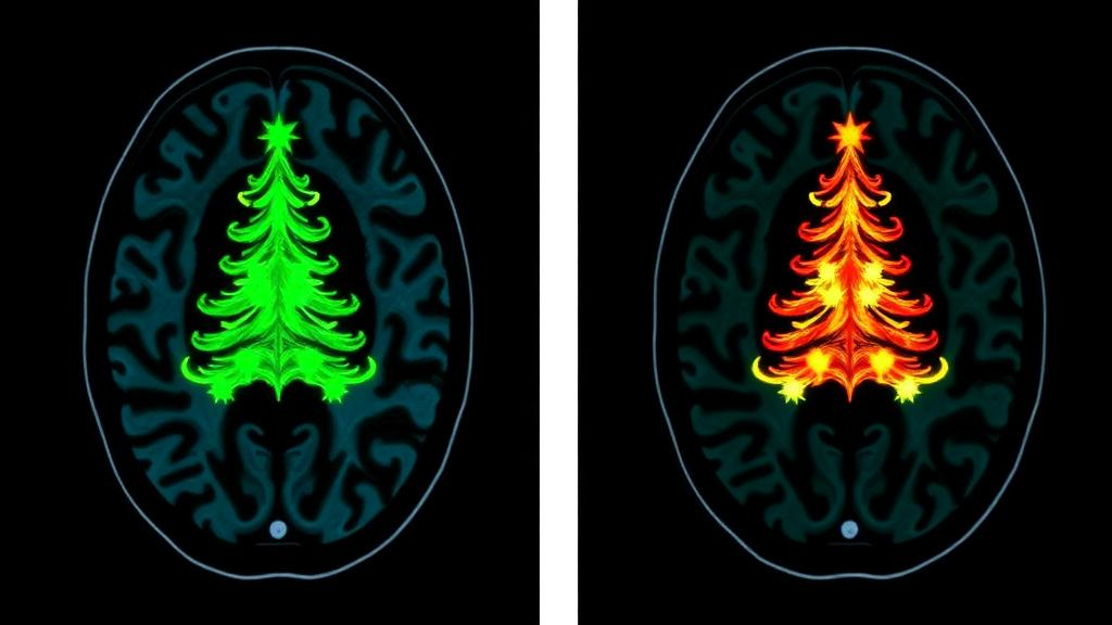
A rare and devastating fungal infection of the central nervous system has been documented in striking detail by radiologists in Hangzhou, China, offering one of the most vivid imaging portraits yet of intracranial talaromycosis. In a case report published in BMC Infectious Diseases, a team at the Second Affiliated Hospital of Zhejiang University School of Medicine describes how an infection with the dimorphic fungus Talaromyces marneffei progressed in a 50-year-old woman to multiple hemorrhagic infarcts, leaving a characteristic fingerprint on magnetic resonance imaging that the authors liken to a Christmas tree. The report, led by Xinyi Chen, Yunyun Jin, Jiahua Wen and corresponding author Biao Jiang, arrives at a moment when clinicians worldwide are being urged to recognize fungal diseases of the brain earlier, because the window for effective treatment is often brutally short.
The clinical story began with a neurosurgical procedure. The patient underwent lysis of spinal arachnoid adhesions as treatment for hydrocephalus, a condition in which cerebrospinal fluid accumulates in the brain’s ventricles and raises intracranial pressure. Postoperatively, rather than recovering as expected, she developed progressive dizziness and limb weakness. Contrast-enhanced magnetic resonance imaging revealed meningitis, inflammation of the membranes that envelop the brain and spinal cord. At that stage, her physicians treated her empirically with piperacillin-tazobactam, a broad-spectrum antibiotic combination, for one month. Antibacterial therapy, however, would prove to be aimed at the wrong class of microbe entirely.
Follow-up imaging changed the picture dramatically. The repeat contrast-enhanced MRI showed striking enhancement of the skull base meninges, arranged in a pattern the authors describe as a vividly characteristic Christmas-tree appearance. In neuroradiology, meningeal enhancement along the skull base is a well-recognized sign of chronic infectious or inflammatory disease, but the branching, tree-like configuration documented here is unusual and visually distinctive. Equally important was a second set of findings: the images demonstrated irregular thickening of cerebral vessel walls that took up contrast material, together with irregularity of the vessel lumens themselves. That combination, vessel wall inflammation plus caliber irregularity, pointed toward a vasculitic process capable of blocking blood flow and causing strokes.
That is precisely what happened. The infection developed into multiple hemorrhagic infarcts, areas of brain tissue that died from interrupted blood supply and then bled into the damaged region. Hemorrhagic transformation of infarcts is a feared complication of any cerebral infarction, and in infectious settings it typically reflects direct invasion or inflammation of vessel walls, which weakens them and disrupts the blood-brain barrier. The imaging sequence in this case therefore traced a coherent pathological arc: meningeal infection, spread to the vessels of the circle of brain perfusion, vasculitis, thrombosis or embolization, infarction, and finally hemorrhage within the infarcted tissue.
The decisive diagnostic step came not from microscopy or culture but from metagenomic next-generation sequencing, or mNGS, performed on the patient’s cerebrospinal fluid and blood samples. mNGS is an unbiased technique that extracts all genetic material in a sample and matches it against databases of known organisms, allowing pathogens that are slow-growing, fastidious, or unexpected to be identified without prior suspicion. The sequencing confirmed infection with Talaromyces marneffei, a thermally dimorphic fungus that grows as a mold at ambient temperature and as a yeast inside the human body. The case underscores how mNGS has transformed the workup of central nervous system infections, where conventional cultures frequently fail and empiric therapy cannot wait.
Talaromyces marneffei, formerly classified as Penicillium marneffei, is endemic to Southeast Asia and southern China, where it resides in environmental reservoirs that include bamboo rats and soil. Historically, human disease occurred almost exclusively in people with advanced immunosuppression, particularly those with untreated HIV infection, in whom talaromycosis was a defining AIDS-related illness. In recent years, however, cases have increasingly been reported in individuals without obvious immune defects, and the fungus has become an emerging concern as HIV-negative talaromycosis is recognized more often. Central nervous system involvement is rare even among infected patients, which is why a fully documented case with serial imaging and molecular confirmation carries outsized educational value for clinicians far beyond the endemic zone.
Treatment was initiated once the diagnosis was established. The patient received liposomal amphotericin B at a dose of 150 milligrams once daily for two weeks, the standard induction approach for severe talaromycosis, which targets fungal cell membranes and is the recommended first-line agent for disseminated disease. Despite this appropriate therapy, her consciousness failed to improve. The likely explanation, implicit in the imaging, is that by the time the correct diagnosis was made, the infection had already inflicted irreversible vascular and parenchymal damage. Fungal vasculitis of the brain is notoriously refractory to antifungal drugs, which penetrate inflamed, thrombosed tissue poorly, and hemorrhagic infarcts do not reverse even when the organism is finally suppressed.
The outcome was grim. The patient was discharged at her family’s request with a poor prognosis. The authors of the report, writing from the Department of Radiology, emphasize the imaging findings themselves as the core contribution: the Christmas-tree pattern of skull base meningeal enhancement, the irregular enhancing vessel wall thickening, and the evolution into multiple hemorrhagic infarcts together form a recognizable syndrome that should raise suspicion for intracranial T. marneffei infection in the right clinical context. Recognizing this pattern matters because it can prompt early mNGS testing of cerebrospinal fluid, compressing the diagnostic delay that so often proves fatal in fungal diseases of the brain.
The case also carries broader lessons about post-neurosurgical infections. The patient’s symptoms emerged after spinal arachnoid adhesion lysis, and while the report does not establish the precise route by which the fungus entered the central nervous system, the temporal association illustrates how iatrogenic disruption of protective barriers can open a door to opportunistic organisms. It also highlights the limits of empiric antibacterial therapy: a month of piperacillin-tazobactam provided no benefit against a fungus, and the delay may have allowed the infection to progress from treatable meningitis to destructive vasculitis. For clinicians managing postoperative meningitis that fails to respond to antibiotics, the report argues for early escalation to molecular diagnostics and antifungal coverage.
Published as an open-access case report and conducted in accordance with the Declaration of Helsinki, with ethics approval from the hospital’s committee and written informed consent from the patient’s next of kin, the study is a small piece of evidence by design, a single patient’s course rather than a trial. Yet in rare diseases, single cases are how the collective clinical eye is trained. As imaging techniques sharpen and sequencing costs fall, reports like this one build the pattern library that allows a radiologist in any country to look at an enhancing skull base, see the branches of a Christmas tree, and know that a treatable diagnosis may still be within reach, if only it is pursued in time.
Subject of Research: Imaging features of intracranial Talaromyces marneffei infection progressing to multiple hemorrhagic cerebral infarcts
Article Title: Imaging findings of intracranial Talaromyces marneffei infection developing into multiple hemorrhagic infarcts: a case report
Article References: Chen, X., Jin, Y., Wen, J., & Jiang, B. (2026). Imaging findings of intracranial Talaromyces marneffei infection developing into multiple hemorrhagic infarcts: a case report. BMC Infectious Diseases. https://doi.org/10.1186/s12879-026-14496-9
Image Credits: AI Generated
DOI: 10.1186/s12879-026-14496-9
Keywords: Talaromyces marneffei, talaromycosis, fungal meningitis, MRI, hemorrhagic infarct, metagenomic next-generation sequencing, cerebrospinal fluid, vasculitis, hydrocephalus, central nervous system infection, amphotericin B, case report
Cite Scienmag News
Ophelia Keating. (October 3, 2026). Rare Fungal Brain Infection Reveals Striking Christmas-Tree Sign on MRI. Scienmag. https://scienmag.com/rare-fungal-brain-infection-reveals-striking-christmas-tree-sign-on-mri/
Ophelia Keating. "Rare Fungal Brain Infection Reveals Striking Christmas-Tree Sign on MRI." Scienmag, 3 October 2026, https://scienmag.com/rare-fungal-brain-infection-reveals-striking-christmas-tree-sign-on-mri/. Accessed 5 October 2026.
Ophelia Keating. "Rare Fungal Brain Infection Reveals Striking Christmas-Tree Sign on MRI." Scienmag. October 3, 2026. https://scienmag.com/rare-fungal-brain-infection-reveals-striking-christmas-tree-sign-on-mri/


