


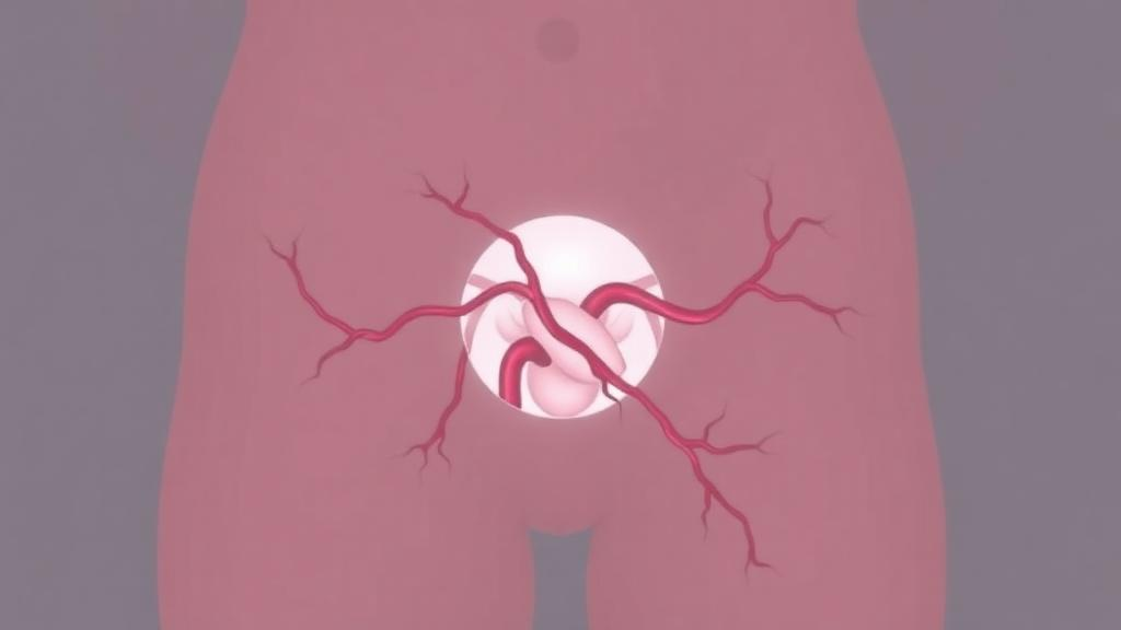
A rare tumor of the pelvic region, so richly supplied with blood vessels that even a simple biopsy triggered difficult bleeding, has been successfully removed after surgeons deployed a strategy borrowed from interventional radiology: cutting off the tumor’s blood supply with tiny detachable coils before making a single surgical incision. The case, reported in the journal Clinical Case Reports, describes how a 22-year-old Japanese woman with a large aggressive angiomyxoma underwent preoperative arterial embolization that allowed her surgical team to excise the entire tumor with an estimated blood loss of just 50 milliliters, a remarkably small amount for a tumor of this size and vascularity.
Aggressive angiomyxoma, often abbreviated AAM, is a mesenchymal tumor that was first described in 1983 and remains exceedingly rare. It occurs predominantly in women of reproductive age and typically arises in the genital tract and the perineal–pelvic region, the soft tissue space between the pelvic floor and the external genitalia. Although the tumor is classified by the World Health Organization as benign and of uncertain differentiation, its behavior is anything but harmless. It grows by locally infiltrating surrounding tissues rather than spreading to distant organs, and it has a stubborn tendency to return after treatment. Reported recurrence rates range from 30 to 50 percent, with most recurrences appearing within three years of surgery, and recurrences have been documented even when pathologists found no tumor cells at the edges of the removed tissue.
The patient in this report presented with irregular genital bleeding and no other relevant medical history. Physical examination revealed a soft, elastic mass extending from her left buttock into the perineal region. Laboratory tests of common tumor markers, including carbohydrate antigen 125 and carbohydrate antigen 19–9, returned values within normal reference ranges, offering few clues about the nature of the mass. Ultrasonography then revealed a well-defined, 12-centimeter tumor with a laminated internal structure and abundant internal blood flow, while the uterus and both ovaries appeared entirely normal, helping to exclude a gynecologic origin.
Magnetic resonance imaging provided the characteristic imaging signature of the tumor. On T2-weighted sequences, the mass showed mixed high and low signal intensities arranged in a swirling, layered pattern, a finding long associated with aggressive angiomyxoma and thought to reflect interlacing strands of tumor tissue within a loose, water-rich matrix. After intravenous contrast administration, the tumor enhanced heterogeneously, confirming its rich vascularity. A positron emission tomography scan using the glucose analog fluorodeoxyglucose showed increased uptake within the mass, and a needle biopsy ultimately yielded the histological diagnosis of aggressive angiomyxoma.
With the diagnosis established, the team planned surgical resection as the definitive treatment. In the roughly one-month interval before the scheduled operation, the patient received oral relugolix at a dose of 40 milligrams once daily. Relugolix is a gonadotropin-releasing hormone antagonist, a drug that suppresses the pituitary signals driving ovarian estrogen production. The rationale rests on a well-documented biological feature of these tumors: the majority express estrogen and progesterone receptors, making them potentially responsive to hormonal manipulation, which has been explored both to shrink tumors before surgery and to control recurrent disease. In this case, however, the strategy did not achieve its goal. No significant change in tumor size was observed during the waiting period, and the operation proceeded as planned.
A decisive clue about the operative risk had already emerged during the diagnostic biopsy. Hemostasis, the process of stopping bleeding, had proven difficult to achieve after the biopsy because of the tumor’s hypervascular nature. Recognizing that significant intraoperative hemorrhage could impede a complete and safe resection, the team turned to preoperative arterial embolization, a minimally invasive procedure in which the arteries feeding a tumor are deliberately occluded. Via an access point in the right femoral artery, the interventional team performed selective angiography, injecting contrast dye to map the tumor’s arterial supply in real time. The images revealed tumor-feeding vessels arising from the left internal pudendal artery, one of the key branches supplying the perineum.
The embolization itself was carried out with detachable coils, soft platinum devices that can be advanced through a catheter, positioned precisely within a target vessel, and released only once the operator confirms optimal placement. The team deployed a total of sixteen coils across two vascular territories. Four coils measuring 2 millimeters by 6 centimeters, six measuring 3 millimeters by 9 centimeters, and two measuring 4 millimeters by 12 centimeters were used to occlude the distal branches of the left internal pudendal artery. Subsequent angiography of the superior and inferior gluteal arteries uncovered a second supply route, with additional feeding vessels branching from the inferior gluteal artery, and these were embolized with six further coils. A final angiographic survey of the left external iliac artery confirmed that no additional feeding vessels remained, and repeat imaging demonstrated near-complete disappearance of tumor blood flow. No complications from the procedure were observed.
Timing mattered. To exploit the window of maximal ischemia before collateral vessels could recruit new flow into the tumor, the surgeons began the operation approximately one and a half hours after the embolization was completed. Working with the patient in the lithotomy position, they made a roughly 10-centimeter incision from the perineum toward the left buttock and circumferentially dissected the tumor free from the surrounding normal tissue. No gross residual tumor was identified after excision, and the operative time was 163 minutes. The estimated blood loss, measured by the gauze-count method, was 50 milliliters, an exceptionally low figure for the resection of a 12-centimeter hypervascular pelvic mass and a striking demonstration of the hemodynamic benefit conferred by the embolization.
The resected specimen displayed the classic gross and microscopic features of the disease. On its outer surface the tumor was soft and smooth, while the cut surface had a gelatinous quality, a reflection of the abundant myxoid, mucus-like matrix that gives the tumor its name. Under the microscope, the lesion consisted of spindle-shaped cells with only mild atypia scattered through a loose myxoid stroma rich in blood vessels. Immunohistochemical staining showed the tumor cells were positive for estrogen receptor, progesterone receptor, smooth muscle actin, and desmin, and negative for CD34, with a Ki-67 proliferation index of just 1 percent, confirming the low proliferative activity typical of this benign neoplasm. The final pathological diagnosis was aggressive angiomyxoma. Because the tumor was dissected along its capsule without taking an additional cuff of surrounding healthy tissue, the pathological margin was positive, designated R1, a common and clinically accepted outcome in the perineal region where wide margins would sacrifice critical structures.
Follow-up has so far been encouraging. Pelvic magnetic resonance imaging performed nine months after surgery showed no evidence of recurrence, and at 18 months the patient remained free of clinical disease. The authors are careful to note the limits of a single case: the efficacy of preoperative embolization for aggressive angiomyxoma cannot be established from one patient, and published experience with this approach remains sparse, including one prior report among 32 pelvic cases in which embolization enabled a complete R0 resection. Still, the case highlights a practical principle. For large or markedly hypervascular tumors, selective angiography both maps the vascular anatomy to guide the surgical approach and reduces intraoperative bleeding, potentially lowering the risk of complications when extensive resection is required or when preservation of reproductive function is a priority. Because the tumor can recur even after long periods of remission, and because this patient’s follow-up spans only a year and a half, long-term surveillance will be essential. The authors call for further accumulation of cases to clarify when preoperative embolization should become a standard part of the surgical planning for this rare and vascularly deceptive tumor.
Subject of Research: Preoperative arterial embolization for surgical resection of hypervascular aggressive angiomyxoma
Article Title: Preoperative Arterial Embolization Facilitating Safe Resection of a Hypervascular Aggressive Angiomyxoma: A Case Report
Article References: Takashima, Y., Terada, S., Tanaka, Y., Yamada, T., Yamamoto, K., Hirose, Y., & Tanaka, T. (2026). Preoperative Arterial Embolization Facilitating Safe Resection of a Hypervascular Aggressive Angiomyxoma: A Case Report. Clinical Case Reports, 14(10), Article e73667. https://doi.org/10.1002/ccr3.73667
Image Credits: AI Generated
DOI: 10.1002/ccr3.73667
Keywords: aggressive angiomyxoma, arterial embolization, detachable coils, pelvic tumor, interventional radiology, surgical resection, GnRH antagonist, relugolix, hypervascular tumor, magnetic resonance imaging, case report, gynecologic surgery
Cite Scienmag News
Ophelia Keating. (October 3, 2026). Coil Embolization Before Surgery Helped Remove a Rare Hypervascular Pelvic Tumor With Minimal Blood Loss. Scienmag. https://scienmag.com/coil-embolization-before-surgery-helped-remove-a-rare-hypervascular-pelvic-tumor-with-minimal-blood-loss/
Ophelia Keating. "Coil Embolization Before Surgery Helped Remove a Rare Hypervascular Pelvic Tumor With Minimal Blood Loss." Scienmag, 3 October 2026, https://scienmag.com/coil-embolization-before-surgery-helped-remove-a-rare-hypervascular-pelvic-tumor-with-minimal-blood-loss/. Accessed 4 October 2026.
Ophelia Keating. "Coil Embolization Before Surgery Helped Remove a Rare Hypervascular Pelvic Tumor With Minimal Blood Loss." Scienmag. October 3, 2026. https://scienmag.com/coil-embolization-before-surgery-helped-remove-a-rare-hypervascular-pelvic-tumor-with-minimal-blood-loss/


