


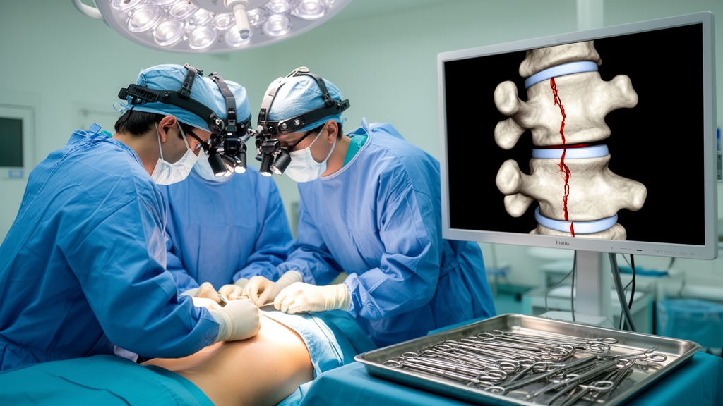
A 42-year-old woman who arrived at a hospital after a road accident turned out to carry one of the rarest lumbar spine injury patterns ever documented: bilateral pars defects at two adjacent vertebrae, L3 and L4, each produced by a completely different mechanism, while the vertebra below, L5, remained entirely intact. The case, published in Clinical Case Reports, describes how surgeons stabilized the unstable segments with pedicle screw fixation and two-level interbody fusion, achieving full neurological recovery within ten weeks.
The pars interarticularis is a narrow segment of bone that connects the superior and inferior articular processes of a vertebra, forming part of the posterior arch. Because it is the thinnest bony bridge in this region, it concentrates mechanical stress during repeated spinal extension and rotation. When loading exceeds the bone’s capacity over time, microscopic damage accumulates and can progress to a complete fatigue fracture, a condition known as spondylolysis. Epidemiological studies cited in the source literature place the prevalence of such defects at roughly four to six percent among athletes and adults exposed to repetitive extension loading, a figure considerably higher than in the general sedentary population. Activities involving frequent hyperextension of the lumbar spine, such as gymnastics, cricket bowling, weightlifting, and diving, are particularly associated with this pathology, which explains why the defect is often discovered in young athletic individuals even before symptoms become disabling.
The natural history of a pars defect is highly variable. Some defects remain asymptomatic throughout life and are discovered incidentally on imaging obtained for unrelated reasons. Others progress to chronic mechanical back pain, and in a subset of patients the complete bony separation allows the vertebral body to slip forward on the vertebra beneath it, producing isthmic spondylolisthesis. Once slippage reaches a significant grade, the narrowed canal and stretched neural structures can produce radicular sensory changes, motor weakness, or, in severe cases, cauda equina symptoms. The Wiltse-Newman classification, referenced in the case, organizes spondylolisthesis by etiology, with Type II covering isthmic defects; subtypes distinguish a fatigue fracture of the pars (Type II A) from elongation or fracture patterns arising through other mechanisms (Type II C). Applying this framework, the treating team labeled the chronic L4 defect as Type II A and the fresh traumatic L3 defect as Type II C, formally acknowledging that two adjacent vertebrae harbored lesions of fundamentally different origin.
The distribution of pars defects across lumbar levels is strongly skewed toward the lower spine. The L5 level accounts for the overwhelming majority of cases, followed by L4, while involvement of L3 is described as very rare. This gradient reflects biomechanics: the lumbosacral junction experiences the greatest shear forces and the greatest range of extension, concentrating fatigue stress at the lowest segments. A defect at L3 therefore stands out as unusual on its own, and the combination of bilateral defects at both L3 and L4, with L5 spared, represents a pattern that the authors could not match in prior reports. Their database search across PubMed, ScienceDirect, and Google Scholar, using terms covering contiguous pars defects, adjacent-level lumbar pathology, and surgical treatment, retrieved only scattered case reports of adjacent-level defects, and those typically involved a single shared mechanism, such as stress fractures in young athletes, rather than two distinct etiologies converging at neighboring levels.
Acute traumatic pars fractures deserve particular attention in the trauma setting. They are uncommon, usually resulting from high-energy mechanisms such as motor vehicle collisions or falls from height, and they can be easily missed when plain radiographs are the primary imaging modality. The pars is a thin structure oriented obliquely on lateral films, and overlying bowel gas, patient positioning, and pain-limited positioning during dynamic views can all obscure a fresh fracture line. In this case, the diagnosis of acuity rested on clinicoradiological correlation: the patient’s two-year history of mechanical low back pain pointed to a pre-existing chronic defect at L4, while the acute high-energy accident, the new neurological deficits in the L3 and L4 dermatomes, and the intraoperative appearance of the L3 defect with sharp, fresh margins supported an acute traumatic origin there. The chronic L4 defect, by contrast, showed fibrotic, sclerotic margins characteristic of a long-standing non-union that had progressed to Grade 2 anterolisthesis.
The absence of preoperative CT and MRI is a notable limitation of the reported management. Cross-sectional imaging would have provided superior characterization of pars morphology, more confident dating of each lesion, direct visualization of neural element compression, and assessment of associated soft-tissue injury such as disc disruption or posterior ligamentous complex damage. Resource constraints at the time of admission made these studies unavailable, and the surgical team relied on dynamic flexion-extension radiographs, the clinical history, and intraoperative findings to differentiate acute from chronic pathology. The authors emphasize that this experience underscores the enduring value of careful clinical assessment and surgical judgment in settings where advanced imaging is not immediately accessible, while acknowledging that CT and MRI remain the standard for preoperative planning whenever they can be obtained.
Neurological findings in this patient merit closer examination. Sensory deficits were confined to the L3 and L4 dermatomes, and motor examination revealed weakness of bilateral hip flexion and knee extension, both functions mediated largely by the L2-L4 nerve roots, graded at 3 out of 5 on the standard Medical Research Council scale. Reflexes were preserved and there was no bladder or bowel involvement, indicating an incomplete injury without cauda equina compromise. This pattern is consistent with compression or irritation of the lumbar nerve roots at the levels of the defects and the Grade 2 slip, rather than a complete structural injury to the spinal cord, which terminates at approximately the L1 level in adults and is therefore not directly involved in lower lumbar fractures.
The surgical decision-making illustrates the tension between two established treatment philosophies for pars defects. Direct pars repair, typically involving fixation of the fractured pars with screws, hooks, or a modified Scott technique, preserves the motion segment and the integrity of the adjacent intervertebral discs, making it attractive for young patients with isolated defects and minimal slippage. Instrumented fusion with pedicle screws and interbody cages, by contrast, sacrifices the fused segments but provides immediate rigid stability, allows thorough neural decompression, and corrects existing deformity. The presence of Grade II spondylolisthesis at L4, neurological deficits requiring decompression, and instability at two contiguous levels tipped the balance decisively toward fusion in this case. A direct repair would not have addressed the slipped segment or the two-level instability, and a shorter construct spanning only one level would have left one of the defective vertebrae inadequately stabilized, particularly after the decompressive laminectomy and facetectomy further weakened the posterior elements.
The operative technique followed standard posterior lumbar fusion principles. Pedicle screws of 6.5 mm diameter and 45 mm length were placed bilaterally at L3, L4, and L5 under fluoroscopic guidance, creating a two-segment construct. Rods were contoured to restore and maintain sagittal lordosis, the natural inward curvature of the lumbar spine whose loss contributes to sagittal imbalance and accelerated adjacent-segment degeneration. Decompression was achieved through laminectomy, removal of the ligamentum flavum, medial parsectomy, and foraminotomy from L3 to L5, widening the neural foramina and relieving pressure on the traversing and exiting nerve roots. At both the L3-L4 and L4-L5 levels, a right-sided facetectomy, cruciate annulotomy, and complete disc excision prepared the intervertebral space, and the end plates were prepared before inserting polyetheretherketone cages packed with autologous bone graft and bone substitute. PEEK cages are favored in interbody fusion for their elastic modulus approximating that of cortical bone and their radiolucent properties, which permit clear assessment of bony consolidation on follow-up imaging. Intraoperative neuromonitoring was used throughout, providing real-time feedback on neural function during instrumentation.
The postoperative course was notably rapid. The patient was mobilized out of bed on the first day after surgery and discharged the following day after drain removal, with drain output of only 100 milliliters and well-controlled pain. Early mobilization after lumbar fusion is increasingly standard practice, supported by evidence that it reduces complications such as deep vein thrombosis, pulmonary atelectasis, and deconditioning without increasing hardware failure rates when the construct is rigid. At the three-week follow-up, the wound had healed satisfactorily, sutures were removed, and the visual analogue scale pain score had fallen from 8 out of 10 to 2 out of 10. Sensation in the affected dermatomes was recovering, and motor strength had improved from 3 out of 5 to a full 5 out of 5. Radiographs at that stage confirmed good alignment and stable hardware positioning.
At approximately ten weeks after surgery, the patient remained neurologically intact, ambulatory, and independent in activities of daily living without complications. Follow-up radiographs demonstrated maintained alignment, stable instrumentation, and progressive fusion across both interbody levels, with no evidence of hardware loosening, implant failure, or loss of correction. The ten-week window corresponds to the early phase of bony consolidation in spinal fusion, during which bridging callus and graft incorporation become radiographically apparent; complete remodeling and solid arthrodesis typically require six to twelve months or longer, so continued surveillance remains appropriate even after this encouraging early result.
Several broader lessons emerge from this report. First, trauma patients presenting with lumbar pain should be asked specifically about prior back symptoms, because a pre-existing spondylolytic defect can fundamentally alter the injury pattern and the treatment plan. Second, atypical injury patterns, such as defects at unusually high lumbar levels or at multiple contiguous levels, warrant careful scrutiny of both acute and chronic contributions. Third, the coexistence of instability, slippage, and neurological deficit generally mandates decompression and instrumented fusion rather than motion-preserving alternatives, even though fusion sacrifices segmental mobility. Finally, the case demonstrates that satisfactory outcomes, including full motor recovery and progressive fusion, can be achieved even when advanced preoperative imaging is unavailable, provided that clinical correlation and intraoperative assessment are rigorous. The authors’ literature review confirms that contiguous bilateral pars defects arising from two distinct mechanisms at L3 and L4, with an intact L5, have not previously been described in the accessible literature, making this a genuinely novel contribution to the documented spectrum of lumbar spine pathology.
Subject of Research: A rare case of contiguous bilateral lumbar pars defects at L3 and L4 from different mechanisms treated with pedicle screw fixation and interbody fusion.
Article Title: Contiguous Lumbar Bilateral Pars Defects at L3 and L4 (With Intact L5): Treated With Pedicle Screw Fixation and Interbody Fusion: A Case Report With Literature Review
Article References: Ali, M. R., Saleem, M. J., Shahid, A., Sajid, M. S., Haris, M., Asmat, S., Afzal, N. U. S., & Al Diab Al Azzawi, M. (2026). Contiguous Lumbar Bilateral Pars Defects at L3 and L4 (With Intact L5 ): Treated With Pedicle Screw Fixation and Interbody Fusion: A Case Report With Literature Review. Clinical Case Reports, 14(10), Article e73556. https://doi.org/10.1002/ccr3.73556
Image Credits: AI Generated
DOI: 10.1002/ccr3.73556
Keywords: lumbar spine, pars interarticularis, spondylolysis, spondylolisthesis, pedicle screw fixation, interbody fusion, spinal trauma, TLIF, case report, spine surgery, biomechanics, neurological recovery
Cite Scienmag News
Ophelia Keating. (October 17, 2026). Rare Double Spine Fracture With Two Different Causes Stuns Surgeons. Scienmag. https://scienmag.com/rare-double-spine-fracture-with-two-different-causes-stuns-surgeons/
Ophelia Keating. "Rare Double Spine Fracture With Two Different Causes Stuns Surgeons." Scienmag, 17 October 2026, https://scienmag.com/rare-double-spine-fracture-with-two-different-causes-stuns-surgeons/. Accessed 17 October 2026.
Ophelia Keating. "Rare Double Spine Fracture With Two Different Causes Stuns Surgeons." Scienmag. October 17, 2026. https://scienmag.com/rare-double-spine-fracture-with-two-different-causes-stuns-surgeons/


