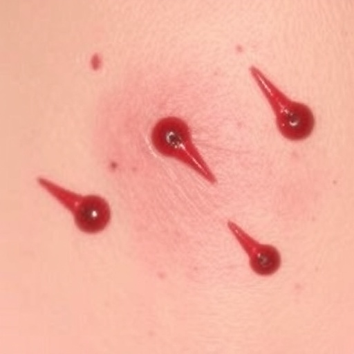
In an extraordinary development within parasitology and infectious disease medicine, researchers have reported a rare and clinically significant case of cutaneous strongyloidiasis manifesting in an immunocompromised patient. The intricate clinical presentation, diagnostic journey, and therapeutic challenges surrounding this infection underscore the complexity of managing parasitic diseases in vulnerable populations. Amid a growing global focus on neglected tropical diseases and immunodeficiency-associated infections, this case sheds critical light on the interplay between host immunity and parasitic pathogenesis, emphasizing the need for heightened vigilance and advanced diagnostic modalities.
Strongyloides stercoralis, the etiological agent in this condition, is a soil-transmitted helminth capable of establishing chronic infections through a complex life cycle involving autoinfection. While strongyloidiasis predominantly affects the intestinal mucosa, cutaneous manifestations are exceedingly rare and often indicative of disseminated or hyperinfection syndrome, especially in immunosuppressed hosts. The reported case diverges from typical presentations and elaborates on an unusual dermatological phenotype, providing valuable clinical insights into the parasite’s behavior and tissue tropism when systemic immune defenses are compromised.
The patient, whose immune status was notably impaired due to underlying medical conditions and possibly immunosuppressive therapy, presented with unique cutaneous lesions. These lesions, initially ambiguous and mimicking other dermatological disorders, prompted a thorough differential diagnostic process. The diagnostic complexity was compounded by the atypical presentation and rarity of cutaneous strongyloidiasis, which traditionally might be overlooked in favor of more common skin infections, leading to potential delays in appropriate treatment.
Advanced diagnostic techniques, including histopathological examination, serological assays, and molecular identification, were pivotal in confirming the presence of Strongyloides larvae within skin biopsy samples. This multifaceted approach highlighted the necessity of integrating conventional parasitology with innovative molecular diagnostic tools, particularly in immunocompromised patients presenting with unusual clinical signs. The utilization of polymerase chain reaction (PCR) amplification of parasite-specific genetic markers facilitated definitive diagnosis, underscoring the significance of molecular diagnostics in contemporary parasitology.
From a pathophysiological perspective, this case elucidates the mechanisms underpinning parasite dissemination beyond the gastrointestinal tract. The aberrant larval migration to cutaneous tissues illustrates the parasite’s adaptive survival strategies in the face of compromised host immunity. Immunosuppression appears to disrupt the containment of autoinfective larvae, permitting widespread tissue invasion and severe clinical manifestations. Understanding these mechanisms enhances clinicians’ ability to anticipate and recognize disseminated infections in susceptible patient groups.
Therapeutically, the management of cutaneous strongyloidiasis demands highly tailored approaches. Anthelmintic regimens, primarily involving ivermectin, demonstrated efficacy in reducing parasitic burden and resolving dermatological symptoms. However, the treatment landscape is complicated by concerns of drug resistance, potential toxicity in immunocompromised patients, and the necessity of prolonged therapy to prevent relapse due to autoinfection cycles. This case emphasizes not only the importance of early intervention but also the critical role of monitoring therapeutic responses through clinical and laboratory parameters.
The immunological interplay elaborated in the case reveals broader implications for understanding host-parasite dynamics. The suppression of cell-mediated immunity, particularly T-cell function, seemingly facilitates parasite persistence and dissemination. This insight reinforces the urgency for clinicians to maintain a high index of suspicion for parasitic infections in patients undergoing immunosuppressive therapies or living with conditions such as HIV/AIDS, hematological malignancies, or undergoing organ transplantation.
Furthermore, the case report stimulates discourse on the epidemiological aspects of strongyloidiasis in non-endemic regions, bringing attention to globalization, migration, and climate change as variables influencing parasite distribution. The emergence of unusual presentations in diverse geographic and demographic contexts challenges existing paradigms and necessitates enhancements in surveillance and public health interventions to mitigate disease burden.
Dermatologists and infectious disease specialists will find this report particularly instructive, illustrating the necessity of multidisciplinary collaboration to accurately diagnose and manage complex parasitic infections. The integration of parasitology expertise with dermatological assessment and immunological evaluation constitutes a model for optimizing patient outcomes in similar scenarios.
In addition to clinical and therapeutic ramifications, this case holds significance for parasitological research. It invites comprehensive studies into the molecular determinants of tissue specificity and host immune evasion by Strongyloides spp. Deciphering these molecular pathways may unlock novel targets for pharmacological intervention and diagnostic innovation, ultimately improving care for patients afflicted with disseminated strongyloidiasis.
From a public health perspective, the report accentuates the importance of screening protocols for strongyloidiasis prior to initiating immunosuppressive regimens. Prophylactic or preemptive treatment could prevent severe cutaneous and systemic manifestations, highlighting preventive strategies as essential components of comprehensive healthcare delivery for at-risk populations.
Moreover, the case underscores the potential for cutaneous lesions to serve as a readily accessible site for diagnosis, potentially facilitating earlier detection than when relying solely on gastrointestinal or pulmonary symptoms. This reorientation towards dermatological signs could inspire a paradigm shift in clinical practice, aiding timely intervention and reducing morbidity.
This patient’s journey through diagnosis and therapy loops back to an essential message: clinicians must maintain a holistic perspective, considering parasitic infections in differential diagnoses even when presentations are atypical. Ignoring such possibilities may lead to fatal outcomes given the parasite’s insidious ability to mimic other diseases and evade routine testing in immunocompromised hosts.
In summary, this rare presentation of cutaneous strongyloidiasis in an immunocompromised patient represents a compelling narrative of a neglected tropical disease intersecting with modern immunopathology. It is a clarion call for the medical community to refine diagnostic algorithms, expand therapeutic armamentaria, and deepen research into parasite-host interactions. The integration of clinical vigilance, advanced diagnostics, and targeted therapy exemplifies the evolving frontier in managing parasitic infections amidst rising immunosuppression worldwide.
As global health landscapes continue to evolve, cases such as this serve as critical reminders of the complexity and adaptability of parasitic pathogens. They highlight the necessity for continuous education, research investment, and multidisciplinary collaboration to confront emerging infectious challenges, ensuring better patient outcomes and advancing the collective understanding of parasitic diseases.
Subject of Research: Rare case of cutaneous strongyloidiasis in an immunocompromised patient, focusing on clinical presentation, diagnostic challenges, immunopathology, and therapeutic management.
Article Title: A Rare Case of Cutaneous Strongyloidiasis in an Immunocompromised Patient: Clinical Insights and Implications.
Article References:
Khanabadi, F., Elmi, T., Didehdar, M. et al. A Rare Case of Cutaneous Strongyloidiasis in an Immunocompromised Patient: Clinical Insights and Implications. Acta Parasit. 70, 165 (2025). https://doi.org/10.1007/s11686-025-01092-1
Image Credits: AI Generated


