


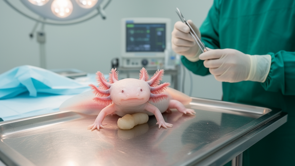
The Mexican axolotl has long been celebrated as biology’s most astonishing survivor, an amphibian that regrows severed limbs, portions of its spinal cord, and even segments of its heart. Now, a nine-year-old female axolotl in Italy has added a new chapter to the species’ medical record: the first documented case of a hepatocellular adenoma, a benign liver tumor, in any urodele amphibian. The case, published in Frontiers in Amphibian and Reptile Science by a team at the University of Parma’s Veterinary Teaching Hospital, describes not only an unprecedented diagnosis but also a remarkably successful treatment and a three-year follow-up that offers fresh hope for owners of these increasingly popular aquatic pets.
The axolotl, a captive-bred female weighing 230 grams and measuring 21 centimeters, arrived at the Exotics Service with a three-week history of progressive swelling of her celomic cavity, the amphibian equivalent of the abdomen. Despite her visibly distended body, she remained alert and responsive to stimuli, a detail that would prove important in guiding her clinicians toward an aggressive diagnostic work-up rather than palliative care. Her husbandry history appeared exemplary, with water maintained at 18 to 19 degrees Celsius, a pH between 6.5 and 8.0, undetectable ammonia and nitrite levels, and a varied diet of earthworms, frozen bloodworms, and defrosted fish offered three times weekly.
The diagnostic odyssey began with radiography in two projections, which confirmed the presence of free fluid, a condition known as hydrocelom or ascites, and revealed a tiny radiopaque focus in the hepatic region. Ultrasonography, performed with a 12 MHz linear probe through a right paramedian ventral approach between the fifth and sixth costal grooves, proved decisive. It demonstrated the free celomic fluid, displaced the internal organs dorsally, identified a 1.2-millimeter gallbladder stone, and, most critically, exposed a mass measuring approximately 1 by 0.7 centimeters on the right liver lobe. Echocardiography showed no cardiac abnormalities, ruling out heart failure as the cause of the fluid accumulation.
Fluid analysis added further clarity. Using a 26-gauge insulin needle under ultrasound guidance, the team aspirated 50 milliliters of clear, odorless fluid from the celomic cavity. Cytological examination identified a pure transudate, a low-protein effusion characteristic of disturbances in fluid regulation rather than inflammation, and found no inflammatory or neoplastic cells. Bacterial culture was negative. A small amount of biliverdin, the green bile pigment typical of amphibians, was noted in the sample, raising suspicion of a cholestatic process related to compression of hepatic tissue by the mass. Blood drawn from the ventral abdominal vein and analyzed within ten minutes showed packed cell volume and serum biochemistry values within established reference ranges for the species.
Computed tomography the following week confirmed the hepatic mass and its dimensions. The animal was sedated with intramuscular alfaxalone at 15 milligrams per kilogram, a catheter was placed in the ventral abdominal vein, and an iodinated contrast agent was administered intravenously at 2 milliliters per kilogram. The entire sedation lasted approximately twenty minutes and was uneventful, a demonstration of how modern anesthetic protocols can be adapted even to a fully aquatic, permanently larval amphibian that breathes primarily through external gills.
Surgery followed within the week. The axolotl was anesthetized by immersion in a buffered tricaine methanesulfonate solution, with the anesthetic water continuously irrigated over her gills by an aquarium pump to maintain oxygenation of the branchial tissue. A pulse oximeter placed on the lingual mucosa and an ECG monitor tracked the depth of anesthesia. Surgeons made a 2.5-centimeter right paramedian incision extending from the fourth to the seventh costal grooves, carefully avoiding the ventral abdominal vein, and used a Lone Star retractor to expose the celomic organs. The liver mass, oval, well demarcated, smooth, and lighter in color than the surrounding parenchyma, was gently elevated with sterile cotton-tipped applicators. Two hemoclips were applied three millimeters from the base of its peduncle before the mass was excised with a No. 11 scalpel blade.
Postoperative care was as meticulous as the procedure itself. The celomic cavity was irrigated with sterile saline at 20 degrees Celsius and inspected for hemorrhage. Muscle layers were closed with absorbable monofilament suture in a simple continuous pattern, and the skin was closed with everted interrupted nylon sutures. Antimicrobial prophylaxis with amikacin was administered every 48 hours for six doses. Recovery took about twenty minutes, with the return of spontaneous movements and gill ventilation as the anesthetic water was gradually replaced. The wound healed without complication, spontaneous feeding resumed ten days after surgery, and skin sutures were removed four weeks postoperatively.
Histopathology delivered the definitive diagnosis. The excised mass was a highly cellular, nodular, non-encapsulated neoplasm compressing the adjacent liver, composed of polygonal hepatocytes with well-defined borders, moderate granular cytoplasm containing coalescing vacuoles, and round basal nuclei with reticular chromatin. Cellular size variation, termed anisocytosis and anisokaryosis, was mild, and not a single mitotic figure was observed across 2.37 square millimeters of tissue. Most tellingly, the tumor lacked portal spaces, the vascular and biliary triads that organize normal liver lobules, a hallmark feature distinguishing hepatocellular adenoma from hepatocellular carcinoma. Histochemical stains for infectious agents, including PAS, Ziehl-Neelsen, and Fite-Faraco, were all negative, and aggregates of melanin-containing cells were noted within the proliferation.
Immunohistochemistry deepened the picture. Using a feline liver sample as a positive control, the team tested the tumor for cytokeratin AE1/AE3, E-cadherin, and vimentin. Cytokeratin-positive cells appeared in biliary epithelium, though less frequently within the neoplastic proliferation than in adjacent parenchyma, while E-cadherin showed membranous expression in both tumor and normal hepatocytes. Vimentin staining was absent, a finding the authors attribute to a lack of cross-reactivity of the RV202 antibody clone compared with the V9 clone used in earlier amphibian tumor studies. These results align with prior immunohistochemical work on hepatocellular and gastric tumors in African clawed frogs, extending the comparative oncology dataset for amphibians in a meaningful way.
The long-term outcome is perhaps the most compelling part of the story. Four months after surgery, the hydrocelom recurred, and the axolotl required periodic drainage of roughly 50 milliliters of pure transudate every two months. Yet serial ultrasonography over the following three years revealed no recurrence of the hepatic tumor or any other celomic lesion, blood analyses remained unremarkable, and the animal maintained a stable body weight of approximately 120 grams, normal spontaneous feeding, and an acceptable quality of life, later developing age-related bilateral cataracts. For a condition in which reported treatment success in amphibians hovers around 36.5 percent, this case suggests that hydrocelom can be managed as a chronic condition rather than a death sentence. It also delivers a clear clinical message: benign hepatic neoplasia belongs on the differential diagnosis list for axolotls presenting with fluid distension, even without overt liver dysfunction, and advanced imaging combined with surgery can deliver years of good-quality life. As captive axolotl populations age and veterinary exotics medicine matures, this landmark case is likely to shape how clinicians worldwide approach swelling in one of the planet’s most endangered and most beloved amphibians.
Subject of Research: First documented case of hepatocellular adenoma and recurrent hydrocelom in a Mexican axolotl, diagnosed with advanced imaging and treated surgically
Article Title: Hepatocellular adenoma associated with recurrent hydrocelom in a Mexican axolotl (Ambystoma mexicanum)- a case report
Article References: Vetere, A., Volta, A., Fumeo, M., Armando, F., Di Pentima, M., Gavezzoli, M., Casalini, J., Di Lecce, R., & Di Ianni, F. (2026). Hepatocellular adenoma associated with recurrent hydrocelom in a Mexican axolotl (Ambystoma mexicanum)- a case report. Frontiers in Amphibian and Reptile Science, 4, Article 1981674. https://doi.org/10.3389/famrs.2026.1981674
Image Credits: AI Generated
DOI: 10.3389/famrs.2026.1981674
Keywords: axolotl, Ambystoma mexicanum, hepatocellular adenoma, hydrocelom, veterinary oncology, amphibian medicine, histopathology, ultrasonography, computed tomography, immunohistochemistry, case report, regenerative medicine
Cite Scienmag News
Nathaniel Bowman. (October 17, 2026). Rare Benign Liver Tumor Removed From Pet Axolotl in Landmark Case. Scienmag. https://scienmag.com/rare-benign-liver-tumor-removed-from-pet-axolotl-in-landmark-case/
Nathaniel Bowman. "Rare Benign Liver Tumor Removed From Pet Axolotl in Landmark Case." Scienmag, 17 October 2026, https://scienmag.com/rare-benign-liver-tumor-removed-from-pet-axolotl-in-landmark-case/. Accessed 17 October 2026.
Nathaniel Bowman. "Rare Benign Liver Tumor Removed From Pet Axolotl in Landmark Case." Scienmag. October 17, 2026. https://scienmag.com/rare-benign-liver-tumor-removed-from-pet-axolotl-in-landmark-case/


