


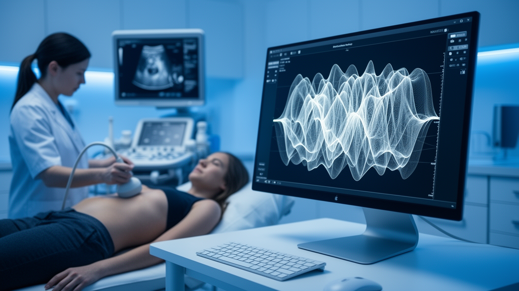
Ultrasound is one of the most trusted tools in modern medicine. Performed tens of millions of times each year worldwide, it avoids ionizing radiation, cuts diagnostic delays, and underpins care in obstetrics, cardiology, and oncology. Yet the safety monitoring that governs every scan still rests on indices conceived decades ago, built on simplified models of tissue that bear little resemblance to the heterogeneous, living organs inside real patients. A new narrative review published in Frontiers in Medical Technology argues that this mismatch is now the central obstacle to truly personalized ultrasound safety, and that a surprising alliance of quantum-inspired algorithms, cloud computing, and edge processors could finally close the gap.
The review, led by Bin Hu and Wei Shi with senior authors Shihong Xiong and Na Gong, examines how the two standard safety parameters, the Mechanical Index (MI) and the Thermal Index (TI), fall short in clinical practice. MI estimates the risk of cavitation, the formation and violent collapse of gas bubbles that can mechanically disrupt tissue. TI models the temperature rise caused by absorbed acoustic energy. Both, however, assume homogeneous tissue. In gas-containing organs, MI can underestimate cavitation risk because it fails to capture nonlinear bubble dynamics. TI, meanwhile, ignores a critical physiological variable: the tissue perfusion rate, or TPR, which determines how quickly blood flow carries heat away from the scanned region.
The authors are refreshingly explicit about what they mean by quantum. No native quantum hardware, no cryogenic qubits, no trapped ions, and no photonic circuits are deployed in any clinical ultrasound suite today. Instead, the quantum layer in their proposed framework refers to quantum-inspired classical algorithms, such as tensor-network compressive sensing and variational-quantum-eigensolver analogues, executed on conventional GPUs and edge processors. These methods emulate quantum parallelism to accelerate multi-parameter inverse problems, and the review cites demonstrations that tensor-network contraction can reproduce the output statistics of quantum-supremacy circuits at polynomial cost, lending the approach benchmark-backed rather than speculative credentials.
The physiological case for dynamic correction is strongest in the kidney. Healthy renal cortex is perfused at more than 4 mL/min/g, generating a robust convective heat sink that attenuates thermal accumulation during prolonged Doppler examinations, while the medulla sits near 0.5 mL/min/g, an order of magnitude lower. A cortex-averaged index therefore systematically underestimates medullary heating precisely where thermal vulnerability is greatest. In transplanted kidneys, where calcineurin-inhibitor vasculopathy already reduces cortical perfusion, the safety margin narrows further, making graft-specific perfusion calibration a first-line requirement rather than an optional refinement.
Perfusion is only half of the story. The review also highlights the acoustic nonlinear parameter B/A, which governs harmonic generation and self-focusing in propagating wavefronts. In soft tissues with elevated B/A, such as fatty liver or fibrotic kidney, acoustic energy transfers from the fundamental frequency into second-harmonic components, improving lateral resolution and contrast-to-noise ratio in experimental phantoms. But the same pathology that boosts harmonic gain simultaneously narrows the cavitation safety margin, and near gas-tissue interfaces or contrast microbubbles, excessive nonlinearity generates cavitation nuclei and standing-wave artifacts. Because B/A rises with fat content and fibrosis, real-time nonlinear mapping becomes a safety necessity, not merely an image-quality garnish.
Crucially, TPR and B/A do not act independently. High perfusion dissipates thermal energy yet simultaneously reshapes local acoustic impedance gradients, modulating nonlinear harmonic generation and cavitation thresholds within the same tissue voxel. A change in TPR therefore propagates nonlinearly into MI estimates via modified B/A profiles, precluding independent parameter correction. This antagonistic coupling creates a non-convex, high-dimensional optimization landscape in which classical gradient descent frequently stagnates in local minima. Tensor-network factorization exploits the low-rank structure of the joint MI-TI-TPR-B/A field, which varies smoothly across connected tissue compartments, to approximate global optima, reducing computational complexity from cubic to near-linear scaling and enabling reconstruction of tissue-specific safety maps within a single cardiac cycle.
The proposed architecture operates through three computational layers. At the edge, on-probe devices perform signal denoising and deep neural network pruning, shrinking data volume before transmission. The streamlined data then flows to the cloud layer, where machine learning algorithms fuse multiple parameters, integrating real-time TPR and acoustic nonlinearity corrections alongside deep learning analysis. Finally, the quantum-inspired layer delivers accelerated optimization and security, with variational solvers minimizing a cost function encoding MI and TI safety constraints, thermal dose limits, and contrast-to-noise ratio metrics, converging within time frames compatible with clinical workflows. During surveillance Doppler of a renal allograft, the edge module would estimate cortical and medullary TPR from a low-MI contrast bolus and map B/A from second-harmonic growth, streaming both fields to the cloud node, where the solver returns the highest-resolution preset whose penalty-weighted safety constraints remain satisfied, all within the same examination.
The qualitative synthesis found that tissue-specific, multi-parameter correction strategies were consistently associated with more conservative acoustic exposure and heightened safety awareness than static MI and TI monitoring alone, although the consistency and clinical magnitude of these benefits varied across anatomical applications and study designs. The authors judged the certainty of the current evidence low to moderate, limited by non-standardized TPR measurement protocols and heterogeneous outcome definitions. TPR estimation currently relies on dynamic contrast-enhanced ultrasound time-intensity curves that suffer from depth-dependent attenuation, inter-vendor software divergence, and the absence of standardized calibration phantoms. Test-retest reproducibility across systems remains unreported for renal applications, and no consensus protocol specifies injection dose, region-of-interest placement, or curve-fitting model.
Data governance adds a further constraint. Cloud-based processing of ultrasound cine loops transmits identifiable patient imaging beyond the scanner, requiring encrypted DICOM transfer, zero-trust access control, and transparent, auditable decision pipelines that current clinical deployments rarely document. Post-quantum cryptography and hybrid quantum-classical key distribution offer a migration path for protecting longitudinal imaging archives against future decryption threats, and lightweight variants are already being adapted to resource-constrained medical IoT devices. Regulatory approval pathways for AI-augmented ultrasound safety monitors remain undefined by major agencies, creating deployment uncertainty that phantom validation studies cannot fully capture.
The review concludes that multi-parameter acoustic correction coupled with cloud computing holds genuine promise for improving ultrasound safety profiling and imaging precision, but that the current low-to-moderate certainty of evidence necessitates future validation of edge-cloud synergies before robust, personalized monitoring systems can be realized. The authors propose a tripartite framework classifying ultrasound safety approaches by adaptive capability, from static monitoring through dynamic correction to quantum-edge integrated real-time adaptation, and identify the kidney, with its steep corticomedullary perfusion gradient and routine Doppler surveillance, as the ideal validation testbed. Near-term progress, they argue, will come from standardized perfusion quantification and validated edge pipelines rather than from quantum acceleration, whose clinical role should be regarded as a long-term, hypothesis-level prospect. For now, the vision of a scanner that knows exactly how much energy your specific tissue can safely absorb remains a compelling hypothesis awaiting rigorous multicenter confirmation.
Subject of Research: ultrasound safety, quantum-inspired computing, tissue perfusion, medical imaging
Article Title: Quantum-edge synergy for dynamic acoustic compensation in ultrasound safety: a narrative review of tissue heterogeneity challenges
Article References: Hu, B., Shi, W., Xiong, S., Li, S., Wang, K., & Gong, N. (2026). Quantum-edge synergy for dynamic acoustic compensation in ultrasound safety: a narrative review of tissue heterogeneity challenges. Frontiers in Medical Technology, 8, Article 1775679. https://doi.org/10.3389/fmedt.2026.1775679
Image Credits: AI Generated
DOI: 10.3389/fmedt.2026.1775679
Keywords: ultrasound, quantum computing, medical imaging, tissue perfusion, cloud computing, patient safety, bioacoustics, Quantum-edge, synergy, dynamic, acoustic, compensation
Cite Scienmag News
Katie Riggs. (October 16, 2026). Quantum-inspired computing may rewrite ultrasound safety rules. Scienmag. https://scienmag.com/quantum-inspired-computing-may-rewrite-ultrasound-safety-rules/
Katie Riggs. "Quantum-inspired computing may rewrite ultrasound safety rules." Scienmag, 16 October 2026, https://scienmag.com/quantum-inspired-computing-may-rewrite-ultrasound-safety-rules/. Accessed 16 October 2026.
Katie Riggs. "Quantum-inspired computing may rewrite ultrasound safety rules." Scienmag. October 16, 2026. https://scienmag.com/quantum-inspired-computing-may-rewrite-ultrasound-safety-rules/


