


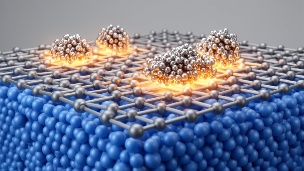
Researchers halted the growth of nickel-doped strontium titanate thin films at less than a single atomic layer and, using in situ scanning tunneling microscopy and spectroscopy, observed nickel cations segregating to the edges of growing islands during the earliest stages of deposition, leaving behind a regular network of pentagonal and hexagonal features roughly 30 nanometers across. This initial fingerprint closely matches the nickel distribution pattern found in thicker films, suggesting the early clusters serve as nuclei for the embedded nickel oxide nanostructures that later channel the exsolution of metallic nickel particles from these materials.
Perovskite oxides with the ABO3 structure are extensively studied and broadly applied, particularly in energy and information technologies, and as epitaxial thin films their crystallinity, orientation, surface morphology, and electrostatic properties can be tailored. Ni-doped SrTiO3 is widely regarded as a typical model system for exsolution, the process in which metal dopants dissolved in a ceramic perovskite lattice migrate to the surface under reducing conditions and emerge as anchored metallic nanoparticles. To synthesize catalysts with a large nanoparticle density, dopant concentrations above 5 atomic percent are typically employed, and co-doping with donor-type dopants is often used to enhance the electrical conductivity of the support oxide. Heavy doping, however, may lead to inhomogeneous cation distributions in ceramic oxides and thin films, giving rise to partial phase separation that is known to be strongly entangled with exsolution behavior. The underlying processes leading to such cation inhomogeneities remain insufficiently discussed, largely because they are difficult to characterize.
The research team grew two closely related systems by pulsed laser deposition on niobium-doped strontium titanate substrates: singly doped SrTi0.95Ni0.05O3-δ, abbreviated STNi, and co-doped SrTi0.9Ni0.05Nb0.05O3-δ, abbreviated STNNi. Growth was monitored layer by layer with reflection high-energy electron diffraction, and the deposition was halted precisely at the first minimum of the RHEED intensity oscillation. At that point, roughly half a monolayer of material had been deposited, leaving isolated perovskite islands standing on an otherwise bare substrate. The samples were transferred under vacuum to a low-temperature scanning tunneling microscope operating at 5 K, allowing the morphology and electronic structure of the nascent film to be probed before a coherent layer ever formed.
Thicker films told the first part of the story. In 150-nanometer-thick specimens examined by scanning transmission electron microscopy with high-angle annular dark-field imaging, the STNi film displayed locally confined bright contrast marking regions of high nickel content, arranged as regular pentagonal or hexagonal features around 30 nanometers in size that linked into a network. At the triple junctions of this network, second-phase NiOx nanocolumns formed, coherently embedded in the perovskite and aligned along the vertical dimension in a manner resembling intentionally fabricated vertically aligned nanostructures, with nickel-rich columns penetrating the full thickness of the film. The co-doped STNNi film, by contrast, showed a denser but less regular pattern of nickel-rich columns extending through the film, with embedded nanostructures that started both from the bottom interface and from intermediate depths, indicating that nucleation begins in the initial growth phase and continues throughout growth.
The sub-monolayer experiments then revealed where those patterns originate. In STM images of the partially covered STNi surface, monolayer-high islands sat on atomically smooth terraces, separated by elongated, linear trenches approximately 20 nanometers in length. Bright clusters exceeding the height of a single unit cell appeared predominantly at the edges of the monolayer islands, right beside the trenches. A watershed algorithm with a height threshold of 4.1 angstroms systematically separated the connected islands, and the resulting regular shapes closely matched the nickel distribution pattern observed in the 150-nanometer film: the mean island area was 494 ± 35 square nanometers for the STM analysis of sub-monolayer islands and 526 ± 39 square nanometers for the transmission electron microscopy analysis of the thick film, corresponding to average diagonals of 27.4 ± 1.1 and 28.3 ± 1.1 nanometers, respectively. The clusters themselves measured 2.5 to 3.5 nanometers in full width at half maximum by STM, consistent with embedded cluster diameters of about 1.5 to 3 nanometers determined by STEM on thicker films, with the slightly larger STM value attributed to tip convolution. In STNNi, the trenches were shorter and less defined, no regular island pattern correlated with the trench locations, and the clusters were much smaller, consistent with more homogeneous and interconnected nucleation.
Spectroscopy added the electronic dimension. On the monolayer islands of STNi, the extracted valence band maximum and conduction band minimum lay 1.8 and 1.5 electron volts from the Fermi level, giving a total bandgap of 3.3 electron volts, consistent with the roughly 3.2 electron-volt bandgap reported for pure strontium titanate. On the phase-separated clusters at the island edges, the valence band edge overlapped with the film spectra, but the conduction band edge shifted 0.4 electron volts upward, widening the total bandgap to 3.7 electron volts. More strikingly, a well-defined in-gap state appeared about 1.2 electron volts above the Fermi level. The researchers assign this state to an acceptor level consistent with the expected role of nickel 3d states in the lattice, and density functional theory calculations support the interpretation: when Ni2+ replaces 12.5 percent of Ti4+ on the B-site, leaving 4.17 percent oxygen vacancies for charge compensation, clear in-gap states appear above the valence band maximum. The calculations cannot unambiguously identify the origin of the observed states, but they indicate that complex defect clusters near NiOx inclusions can plausibly produce them. The enlarged bandgap also matches the reported 3.65 to 3.88 electron-volt bandgap of nickel oxide, and fast Fourier transform analysis confirmed rocksalt NiOx inclusions in both film types, suggesting the cluster spectra represent a convolution of NiOx and defect-cluster contributions.
The co-doped film behaved entirely differently. In STNNi, the monolayer showed a comparable bandgap of about 3.4 electron volts, but the clusters had a very similar fitted bandgap to the surrounding monolayer, and no distinct in-gap peaks appeared. Instead, the spectra showed a shoulder at the valence band maximum and a slight shift toward n-type character, a signature the authors attribute to the donor niobium doping. The absence of in-gap signatures may be related to co-compensation of the niobium and nickel dopants, which would lower the concentration of the oxygen vacancies needed to form the suspected defect clusters. The researchers propose that adding niobium stabilizes nickel in the perovskite lattice and limits its segregation to island edges, an idea supported by prior suggestions that compensating acceptor-donor co-doping can increase dopant solubility in complex oxides. From the perspective of ionic radii, Ni(II) is larger than Nb(V), which in turn is larger than Ti(IV), so incorporating both smaller and larger dopants may help stabilize the structure and suppress nanocolumn formation, yielding the more homogeneous morphology observed in STNNi.
The mechanism the team proposes is elegantly simple. During initial sub-monolayer growth, nickel cations preferentially segregate to the edges of the STNi islands. As deposition continues and coverage increases, the monolayer islands merge into a coherent film, and at the triple junctions between islands enough nickel concentrates that defect clusters form and a NiOx second phase nucleates. The interconnected networks of nickel-rich strontium titanate seen in thick films are thus residues of that early segregation, a literal fingerprint of the growth islands. Because the phase separation occurs on top of each atomic step terrace, terrace width, step size, or step bunching on larger scales does not influence the observed behavior.
To connect the initial pattern to functional behavior, the researchers reduced the sub-monolayer STNi by annealing in ultrahigh vacuum. Annealing at 400 degrees Celsius for 20 minutes produced no significant changes in surface morphology or electronic structure. But at 600 degrees Celsius for 10 minutes, the surface transformed: the trench structure that had formed the pentagonal and hexagonal patterns became less pronounced as islands coalesced, and brighter spots corresponding to exsolved particles emerged with enhanced contrast, with diameters ranging from 3.5 to 6.5 nanometers. Watershed analysis determined a particle coverage of 1.1 percent of the surface area, with the caveat that tip convolution broadens the apparent features. Spectra acquired from the exsolved particles showed metallic behavior and distinct oscillations at approximately +0.35 and +1.1 volts within the conduction band, which the team interprets as a quantum well phenomenon resulting from confinement in the metallic nanoparticles; assigning the first quantized energy level to +0.35 electron volts yields an estimated quantum dot diameter of about 5 nanometers, in good agreement with the observed particle sizes.
The quantitative bookkeeping tied the two reservoirs of nickel together. Assuming semi-ellipsoidal particles with an average height of about 4 angstroms, the total number of exsolved nickel atoms in a representative 200 by 200 square-nanometer area came to approximately 10,000, closely matching the theoretical expectation of about 10,000 atoms for a 5 percent Ni-doped film with 75 percent sub-monolayer coverage. Nearly all of the initially deposited nickel ions therefore accumulated as surface nanoparticles after reduction, indicating full nickel diffusion to the surface, consistent with bulk mass transport posing little constraint in a monolayer-thick sample. Comparing the nickel contained in the as-grown clusters with the total, the researchers estimated an upper limit of roughly 2,000 nickel atoms initially present as distinct nanoclusters, corresponding to about 20 percent of the nominal nickel content, while the remaining approximately 80 percent resided within the perovskite lattice. Both reservoirs contributed to exsolution, demonstrating parallel pathways from pre-nucleated clusters and from the pristine lattice. Beyond clarifying how heavily doped strontium titanate organizes itself from its very first atomic layer, the findings underscore the importance of understanding phase separation from the earliest stages of film growth and offer guidelines for designing functional oxide thin films with tailored properties for catalysis, electronics, and energy devices.
Subject of Research: Initial-stage phase separation and nickel exsolution in doped strontium titanate thin films
Article Title: Partial Phase‐Separation in the Initial Growth Stage of Exsolution‐Active SrTi0.95‐xNi0.05NbxO3‐δ Thin Films
Article References: Liu, Y.-P., Weber, M. L., Jennings, D., Rusevich, L. L., Kotomin, E., Timm, R., Gunkel, F., & Dittmann, R. (2026). Partial Phase‐Separation in the Initial Growth Stage of Exsolution‐Active SrTi 0.95‐x Ni 0.05 Nb x O 3‐δ Thin Films. Advanced Science, 13(59), Article e76812. https://doi.org/10.1002/advs.76812
Image Credits: AI Generated
DOI: 10.1002/advs.76812
Keywords: exsolution, strontium titanate, perovskite oxides, thin films, phase separation, scanning tunneling microscopy, pulsed laser deposition, nickel oxide nanocolumns, dopant segregation, scanning transmission electron microscopy, density functional theory, Partial
Cite Scienmag News
Denise Maddox. (October 25, 2026). Phase Separation in Exsolution Catalysts Begins in the First Atomic Layer. Scienmag. https://scienmag.com/phase-separation-in-exsolution-catalysts-begins-in-the-first-atomic-layer/
Denise Maddox. "Phase Separation in Exsolution Catalysts Begins in the First Atomic Layer." Scienmag, 25 October 2026, https://scienmag.com/phase-separation-in-exsolution-catalysts-begins-in-the-first-atomic-layer/. Accessed 25 October 2026.
Denise Maddox. "Phase Separation in Exsolution Catalysts Begins in the First Atomic Layer." Scienmag. October 25, 2026. https://scienmag.com/phase-separation-in-exsolution-catalysts-begins-in-the-first-atomic-layer/


