


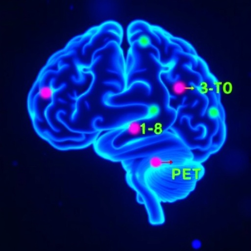
A new PET imaging approach is poised to change how scientists measure gene activity in the living brain. In a study published in Nature Biomedical Engineering, researchers report a purpose-built PET reporter ligand designed to quantify gene expression with far greater precision than earlier, more indirect methods. The work targets a long-standing gap in neurobiology: while gene activity can be traced using molecular tools in cells, translating that information into quantitative, whole-brain measurements in humans and animals has been difficult.
At the core of the advance is the concept of a reporter ligand that behaves like a molecular readout. Rather than simply indicating where cells are, the ligand provides a radioactive signal linked to gene expression dynamics. When the brain expresses a specific target genetic program, the compound’s binding and resulting PET signal reflect that activity, enabling investigators to move beyond “presence or absence” toward actual measurement of expression levels.
The team emphasizes quantitative imaging, a requirement for comparing gene regulation across time, brain regions, or treatment conditions. PET, with its sensitivity to low concentrations of radiotracers, offers a natural platform for this task—but only if the tracer’s chemistry and biological specificity are tuned to report gene-dependent processes reliably. The study therefore focuses on rigorous characterization of how the ligand distributes in neural tissue and how its signal correlates with the underlying genetic output.
Technically, the tracer’s performance depends on multiple factors: specificity for the reporter context, stability in vivo, and favorable pharmacokinetics so that PET can capture meaningful changes rather than background fluctuations. The researchers also discuss how imaging parameters and temporal sampling can be optimized to extract quantitative parameters, such as standardized uptake and other model-based readouts derived from the PET signal.
Importantly, the researchers frame the ligand as a scalable tool for mapping gene expression across the brain. This could support studies of neurological disease mechanisms where gene regulation shifts subtly but meaningfully, such as in neuroinflammation, synaptic dysfunction, or tumor biology. Because PET can be repeated in the same subject, longitudinal studies become feasible, allowing scientists to track gene activity as conditions evolve or therapies are administered.
While gene-reporting systems have existed in preclinical settings, PET offers a unique advantage: whole-organ, noninvasive visualization in vivo. By bridging molecular genetics and clinical imaging technology, the new reporter ligand advances a pathway toward translation, including potential future applications where patients might be monitored for gene expression changes without repeated biopsies.
Overall, the study highlights an engineering principle that is likely to influence the next generation of molecular imaging: the tracer must be tailored to the biology it reports. With that alignment, PET can become not just a detector of molecular targets, but a quantitative instrument for gene regulation in the brain.
Cite Scienmag News
Juliet Wilcox. (July 27, 2026). PET Ligand Enables Quantitative Imaging of Brain Gene Expression. Scienmag. https://scienmag.com/pet-ligand-enables-quantitative-imaging-of-brain-gene-expression/
Juliet Wilcox. "PET Ligand Enables Quantitative Imaging of Brain Gene Expression." Scienmag, 27 July 2026, https://scienmag.com/pet-ligand-enables-quantitative-imaging-of-brain-gene-expression/. Accessed 4 September 2026.
Juliet Wilcox. "PET Ligand Enables Quantitative Imaging of Brain Gene Expression." Scienmag. July 27, 2026. https://scienmag.com/pet-ligand-enables-quantitative-imaging-of-brain-gene-expression/


