


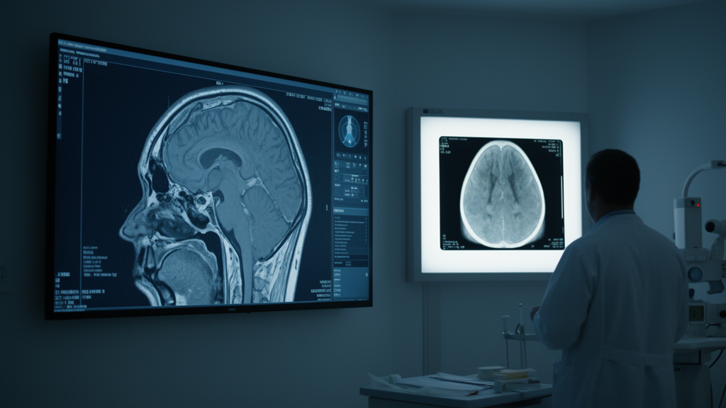
Orbital imaging has traveled a remarkable arc over the past century, moving from plain radiographs that could only hint at disease behind the eye to computed tomography, magnetic resonance imaging, and newer techniques that characterize tissue activity and vascular flow. A new editorial in Frontiers in Ophthalmology, published in the section on Oculoplastics, Orbit and Trauma, frames this evolution and gathers seven contributions into a Research Topic spanning thyroid eye disease, orbital masses, vascular anomalies, lacrimal gland disease, and ocular surface neoplasia. The editorial, authored by Alon Kahana of Oakland University William Beaumont School of Medicine, emphasizes a central theme that runs through the collected work: diagnostic value depends on matching the imaging modality to the specific clinical question.
Choosing the right tool, the editorial argues, matters as much as the technology itself. A second recurring message concerns standardization. Measurements of orbital structures, muscle enlargement, and lesion dimensions are only comparable across patients and time points if they are acquired and reported consistently.
The contributions collected in the Research Topic underscore that interpreting findings in clinical context is equally essential, since the same image can carry different implications depending on the patient's presentation and disease course. The remaining contributions extend across the wider orbit. The editorial notes that techniques capable of characterizing tissue activity and vascular flow are expanding what orbital imaging can contribute to these questions.
As an editorial introducing a Research Topic, the article is a synthesis rather than a single experimental study, and its conclusions reflect the scope of the seven included contributions. Even so, its framing is clear: the future of orbital imaging lies not in any single technology but in disciplined, question-driven use of the full toolkit, from radiography's historical foundations to modern MRI and three-dimensional techniques that continue to reshape diagnosis and care in orbital disease.
Subject of Research: Advances and challenges in orbital imaging for orbital and ocular disease
Article Title: Editorial: Advances and challenges in orbital imaging: from radiography to modern MRI and 3D technologies
Article References: Kahana, A. (2026). Editorial: Advances and challenges in orbital imaging: from radiography to modern MRI and 3D technologies. Frontiers in Ophthalmology, 6, Article 2005013. https://doi.org/10.3389/fopht.2026.2005013
Image Credits: AI Generated
DOI: 10.3389/fopht.2026.2005013
Keywords: orbital imaging, computed tomography, magnetic resonance imaging, thyroid eye disease, orbital masses, vascular malformations, lacrimal gland disease, ocular surface squamous neoplasia, oculoplastics, 3D imaging, Editorial, Advances
Cite Scienmag News
Ophelia Keating. (October 27, 2026). Orbital Imaging Evolves From Radiographs to MRI and 3D Techniques. Scienmag. https://scienmag.com/orbital-imaging-evolves-from-radiographs-to-mri-and-3d-techniques/
Ophelia Keating. "Orbital Imaging Evolves From Radiographs to MRI and 3D Techniques." Scienmag, 27 October 2026, https://scienmag.com/orbital-imaging-evolves-from-radiographs-to-mri-and-3d-techniques/. Accessed 27 October 2026.
Ophelia Keating. "Orbital Imaging Evolves From Radiographs to MRI and 3D Techniques." Scienmag. October 27, 2026. https://scienmag.com/orbital-imaging-evolves-from-radiographs-to-mri-and-3d-techniques/


