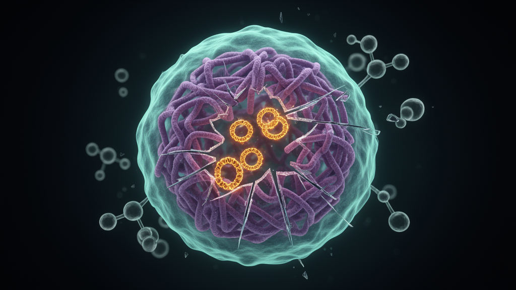
Cancer genomes are notorious for their chaos, but some of their most dangerous weapons are not where they should be. Oncogenes such as MYC, EGFR and CDK4 can break free from their chromosomes and multiply on tiny circular DNA molecules called extrachromosomal DNA, or ecDNA, which float inside the nucleus and drive aggressive tumor growth. A team led by researchers at the University of California, San Diego, and Stanford University has now unveiled AmpliconSuite, an end-to-end computational toolkit published in Nature Methods that identifies these rogue amplifications from standard cancer whole-genome sequencing data, classifies their formation mechanisms, and shares results through a community repository hosting more than 15,000 publicly available samples.
Focal amplifications, meaning concentrated copies of oncogenes on short stretches of the genome, are among the most frequent events in cancer, yet different amplification modes carry very different consequences. ecDNA-mediated amplification is associated with exceptionally high transcription, rapid copy-number adaptation and therapeutic resistance, while breakage–fusion–bridge cycles and homogeneously staining regions follow more conventional chromosomal routes. Distinguishing these modes from sequencing data alone has long been a bioinformatic challenge, because the structural variants and copy-number patterns of each mechanism overlap in confusing ways. AmpliconSuite was designed to resolve exactly this ambiguity.
At the core of the toolkit sits AmpliconArchitect, an algorithm that jointly analyzes structural variants and copy numbers from whole-genome sequencing to reconstruct the architecture of amplified regions. The new AmpliconSuite-pipeline wraps this engine into a complete workflow: users provide sequencing reads in FASTQ, BAM or CRAM format, the pipeline optionally performs alignment and genome-wide copy-number calling with CNVkit to identify candidate focal amplification regions, and AmpliconArchitect then constructs a copy-number-aware breakpoint graph for each candidate. A companion module, AmpliconClassifier, applies rule-based criteria to label every amplification as ecDNA, breakage–fusion–bridge, complex noncyclic or linear amplification. The pipeline is available through Bioconda and Nextflow, making it accessible to any laboratory with sequencing data.
The team substantially improved AmpliconArchitect’s performance since its original release. Version 1.5.r2 achieved a median speedup of 2.8 times over the 2019 version, rising to 6.4 times for samples that originally took more than two hours to process. Structural variant detection was sharpened through entropy filtering of breakpoint-end sequence content and stricter mapping quality requirements, reducing false positives from low-complexity regions. The pipeline also detects low properly-paired read rates and adjusts parameters to suppress artifactual structural variants, while supporting user-provided variant files to augment discovery. Reference genomes for GRCh38, mouse mm10 and even a viral-augmented GRCh38 build are now supported.
Validation was rigorous. The researchers assembled a metaphase fluorescence in situ hybridization dataset covering 175 probed genes across 72 cancer cell lines with closely matching whole-genome sequencing, including 56 ecDNA-borne genes, 52 non-native homogeneously staining regions and 39 native regions. AmpliconClassifier version 1.5.1 achieved a precision of 0.91 and a recall of 0.95 on this benchmark, improving over previous versions from 2020 and earlier. Breakage–fusion–bridge detection was measured against an optical genome mapping dataset of 93 previously analyzed candidates, yielding a recall of 0.71, specificity of 0.95 and precision of 0.88. In silico dilution experiments mixing cancer cell line reads with normal diploid reads across purity levels from 4.8 to 95.2 percent recovered 79.5 percent of theoretically detectable ecDNA species.
A particularly subtle problem the team confronted is that ecDNA does not always stay extrachromosomal. Complete integration of ecDNA into chromosomes is common in cancer cell lines, typically appearing as a non-native homogeneously staining region, meaning an amplified gene sitting on a chromosome other than its chromosome of origin. In the glioma cell line CCF-STTG1, sequencing identified a single CDK6 ecDNA cycle fully explaining the amplification, yet three of nineteen metaphase images also showed non-native CDK6 staining regions, indicating that ecDNA-derived HSRs may coexist latently with the circular molecules. The researchers therefore marked FISH:non-native HSR cases with sequencing-based ecDNA classification as neutral in performance calculations, acknowledging the limits of bulk sequencing in resolving these mixed states.
To democratize data sharing, the team built AmpliconRepository, a Django-based web platform where users create public or private projects from uploaded AmpliconSuite-pipeline outputs. Project pages host overviews of focal amplification abundance, sample pages display interactive copy-number profiles built with Plotly and IGV, and a site-wide search enables custom batch downloads. Unlike other ecDNA databases, AmpliconRepository provides complete tool outputs and lets contributors directly create, update and expand their projects. The repository currently hosts results from more than 15,000 samples, including pan-cancer datasets spanning ICGC, TCGA, PCAWG, HMF and GLASS cohorts.
Applying the toolkit to 6,506 patient samples across these cohorts revealed striking patterns in ecDNA-mediated oncogene co-amplification. AmpliconClassifier identified 2,413 predicted ecDNA events, with 36 percent of ecDNA-positive samples carrying multiple ecDNA. Across the pan-cancer data, ecDNA carried 1,664 oncogenes across 5,348 total megabase pairs, 2.5 times the oncogene density found in the GRCh37 reference genome. The team hypothesized that gene pairs frequently co-amplified on ecDNA would reveal selective pressures shaping ecDNA evolution, and implemented a queryable co-amplification explorer on AmpliconRepository to investigate.
The CDK4-MDM2 co-amplification, observed in 61 samples, was predominantly ecDNA driven: 64 percent by multi-interval ecDNA, 8 percent by different ecDNA, 18 percent with one gene on ecDNA and the other by chromosomal amplification, and only 10 percent by chromosomal amplifications alone. Across chromosome 12q14-15, ecDNA typically excluded the intergenic region between CDK4 and MDM2, yet the chromatin remodeling factor YEATS4, located 514 kilobase pairs downstream of MDM2, was retained alongside MDM2 in 79 percent of MDM2-ecDNA samples. Conversely, the upstream immunomodulatory gene IFNG showed lower co-inclusion at 38 percent, suggesting context-dependent pressures shape ecDNA co-amplification partners. On chromosome 11q13, CCND1 was a frequent chromosomal amplification target, while 57 percent of focal amplifications covering a smaller subregion containing PAK1, RSF1, INTS4 and GAB2 amplified through ecDNA, with 66 percent of those amplifying all four genes together. The most frequent multi-chromosomal co-amplifications paired chromosome 8p12 and 11q13 oncogenes, preferentially via ecDNA, combining signaling oncogenes such as FGFR1 and PAK1 with chromatin remodeling factors RSF1 and NSD3.
To classify ecDNA formation mechanisms, the team developed ecContext, a hierarchical decision tree that categorizes ecDNA into seven structural classes based on copy-number states, transitions, crossing structural variants and foldback inversions. Simple excisional categories, including simple circular with simple background and two-foldback, exhibit fewer copy-number states and a dominant heaviest cycle, while chromothripsis-associated categories, including heavily rearranged unichromosomal and multichromosomal classes, show extensive breakpoint reuse. Validation against experimentally induced ecDNA from Cre-loxP excision models and methotrexate-treated cells demonstrated concordance between predicted and observed mechanisms, with ecContext correctly labeling all 11 excisional ecDNA as simple circular and 87 percent of chromothripsis-derived ecDNA receiving chromothripsis-associated labels. Applied to pan-cancer data, ecContext revealed that 36.8 percent of ecDNA were simple excisional, 48.4 percent were chromothripsis associated and 14.8 percent had unclear origin. Prostate and brain cancers showed enrichment for simple excision-like ecDNA, while skin cancers and sarcomas showed more chromothripsis-associated ecDNA, suggesting tissue-specific factors influence ecDNA formation. The team notes that ecDNA enables rapid modulation of copy numbers, potentially contributing to therapeutic resistance, bridging distant genomic regions and selectively incorporating chromatin remodelers, and that identifying ecDNA-competent cells is biologically and clinically informative as ecDNA-bearing subclones may persist and reselect under selective pressure.
Subject of Research: AmpliconSuite: an end-to-end workflow for analyzing focal amplifications in cancer genomes
Article Title: AmpliconSuite: an end-to-end workflow for analyzing focal amplifications in cancer genomes
Article References: Luebeck, J., Liefeld, T., Wong, I. T.-L., Huang, E., Kim, F., Dameracharla, B., Chan, M., Ahuja, R., Fetter, K., Khatri, D., Zhu, K., Tabor, T., Schreyer, D., Prasad, G., Gnanasekar, A., Raeisi Dehkordi, S., Spandau, S., Adamaszek, M., Kenkre, R., … Bafna, V. (2026). AmpliconSuite: an end-to-end workflow for analyzing focal amplifications in cancer genomes. Nature Methods. https://doi.org/10.1038/s41592-026-03264-x
Image Credits: AI Generated
DOI: 10.1038/s41592-026-03264-x
Keywords: AmpliconSuite, end-to-end, workflow, analyzing, focal, amplifications, cancer, genomes, scientific research
Cite Scienmag News
Nathaniel Bowman. (October 16, 2026). New toolkit decodes how cancer genes shatter, circle and resist. Scienmag. https://scienmag.com/new-toolkit-decodes-how-cancer-genes-shatter-circle-and-resist/
Nathaniel Bowman. "New toolkit decodes how cancer genes shatter, circle and resist." Scienmag, 16 October 2026, https://scienmag.com/new-toolkit-decodes-how-cancer-genes-shatter-circle-and-resist/. Accessed 16 October 2026.
Nathaniel Bowman. "New toolkit decodes how cancer genes shatter, circle and resist." Scienmag. October 16, 2026. https://scienmag.com/new-toolkit-decodes-how-cancer-genes-shatter-circle-and-resist/