A groundbreaking new study from National Jewish Health has unveiled critical insights into sarcoidosis, a complex inflammatory disease notoriously difficult to categorize due to its variable impact on lung tissue. Published in the prestigious journal CHEST, this research illuminates how distinct patterns seen on high-resolution CT scans correspond to the severity and physiological disruptions caused by sarcoidosis in the lungs. It represents a significant leap forward in understanding the disease’s heterogeneous nature and tailoring more precise clinical interventions.
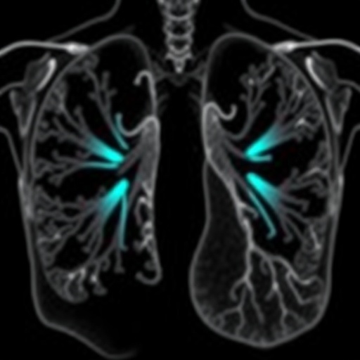
Sarcoidosis, which predominantly involves inflammatory granulomas in lung tissue, presents a clinical spectrum ranging from mild symptoms to severe, progressive lung damage. Central to the study’s inquiry was the distinction between patients exhibiting fibrotic, or scarred, lung abnormalities versus those with nonfibrotic changes or no detectable damage on advanced imaging. Fibrosis denotes a replacement of normal lung architecture with stiff, scar tissue, profoundly compromising the organ’s respiratory function.
The research cohort consisted of more than 900 individuals, facilitating a robust epidemiological and clinical analysis. By integrating high-resolution computed tomography (HRCT) imaging data with comprehensive pulmonary function testing, researchers succeeded in stratifying patients into three discrete categories. Those classified with fibrotic sarcoidosis tended to be older, had endured the disease for longer durations, and exhibited markedly impaired lung function metrics, including reductions in forced vital capacity and diffusion capacity. In stark contrast, patients without fibrosis showed relatively preserved lung function, underscoring the heterogeneity of sarcoidosis pathology.
Remarkably, nearly two-thirds of subjects with fibrotic lung disease demonstrated abnormal pulmonary function tests indicative of restrictive and sometimes combined obstructive deficits. These impairments were less prevalent among those without fibrotic changes. This differential functional compromise suggests that fibrotic progression in sarcoidosis is a critical determinant of respiratory disability, with potential implications for prognostication and management.
Dr. Lisa Maier, a leading authority in environmental and occupational health and co-senior author of the paper, emphasized that sarcoidosis is not a monolithic disease. By meticulously analyzing CT imaging patterns, clinicians can now better identify subpopulations at higher risk of severe lung impairment. Such stratification is invaluable for designing personalized monitoring regimens and therapeutic strategies tailored to disease subtype rather than treating sarcoidosis as a homogeneous entity.
Further granularity was achieved by characterizing specific fibrotic patterns. Patients exhibiting large conglomerate masses — extensive clusters of fibrotic tissue coalescing on imaging — were found to have more complicated lung function impairments. They frequently demonstrated a combination of restrictive and obstructive deficits, a phenotype associated with more significant clinical challenges compared to simpler fibrotic patterns. These imaging biomarkers thus hold promise as surrogate indicators of disease severity and complexity.
Dr. David Lynch, an expert radiologist involved in the study, highlighted that linking radiographic phenotypes with physiological metrics offers a pivotal bridge between diagnostic imaging and patient-centric outcomes. This correlation enhances clinicians’ ability to interpret what is visually discernible on scans with a nuanced understanding of the functional impact of sarcoidosis on individual patients’ respiratory health.
Importantly, a substantial subset of patients bearing no fibrotic changes on CT scans maintained near-normal lung function, a testament to the disease’s variable course. This heterogeneity challenges the one-size-fits-all clinical approach often employed in sarcoidosis management and underscores the pressing need for refined diagnostic frameworks that incorporate both morphological and functional parameters.
Currently, clinical guidelines for pulmonary sarcoidosis do not mandate CT scanning for routine evaluation, nor do they differentiate therapeutic pathways based on fibrotic status. This study’s revelations strongly advocate for integrating advanced imaging into standard clinical protocols to facilitate more accurate disease classification, prognosis, and individualized treatment algorithms.
The research team envisions that this paradigm — combining lung imaging phenotypes with detailed physiological profiling—will catalyze future investigation aimed at earlier identification of patients at risk for severe disease progression. Such prognostic clarity has the potential to usher in novel therapeutic regimens designed to prevent or mitigate fibrotic evolution in sarcoidosis.
As a recognized Center of Excellence by the World Association of Sarcoidosis and Other Granulomatous Disorders (WASOG), National Jewish Health continues to spearhead comprehensive translational research bridging fundamental science and clinical care. Their efforts have culminated in major advances in understanding sarcoidosis pathobiology and enhancing patient outcomes worldwide.
This pivotal study dramatically redefines how clinicians perceive sarcoidosis subtypes and paves the way for personalized medicine strategies in a disease historically marred by diagnostic ambiguity and treatment challenges. By elucidating the nuanced interplay between imaging features and lung function, this investigation effectively charts a course toward more effective, tailored therapeutic interventions for sarcoidosis patients globally.
Subject of Research: Demographic and physiological differences in fibrotic versus nonfibrotic CT imaging subtypes of sarcoidosis and their relationship to pulmonary function impairment.
Article Title: Demographic and Physiological Differences Between Fibrotic and Nonfibrotic CT Subtypes of Sarcoidosis
News Publication Date: 5-Mar-2026
Web References:
https://journal.chestnet.org/article/S0012-3692(26)00270-9/fulltext
http://dx.doi.org/10.1016/j.chest.2026.02.006
Keywords: Sarcoidosis, pulmonary fibrosis, high-resolution CT, lung imaging, pulmonary function testing, granulomatous disease, restrictive lung disease, obstructive lung disease, radiologic phenotypes, personalized medicine
